6QN3

Structure of the Glutamine II Riboswitch


Experimental Data Snapshot

  • Method: X-RAY DIFFRACTION
  • Resolution: 2.30 Å
  • R-Value Free: 0.294 
  • R-Value Work: 0.242 
  • R-Value Observed: 0.245 

wwPDB Validation   3D Report Full Report


This is version 1.3 of the entry. See complete history


Literature

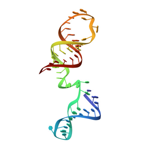
Structure and ligand binding of the glutamine-II riboswitch.

Huang, L.Wang, J.Watkins, A.M.Das, R.Lilley, D.M.J.

(2019) Nucleic Acids Res 47: 7666-7675

  • DOI: https://doi.org/10.1093/nar/gkz539
  • Primary Citation of Related Structures:  
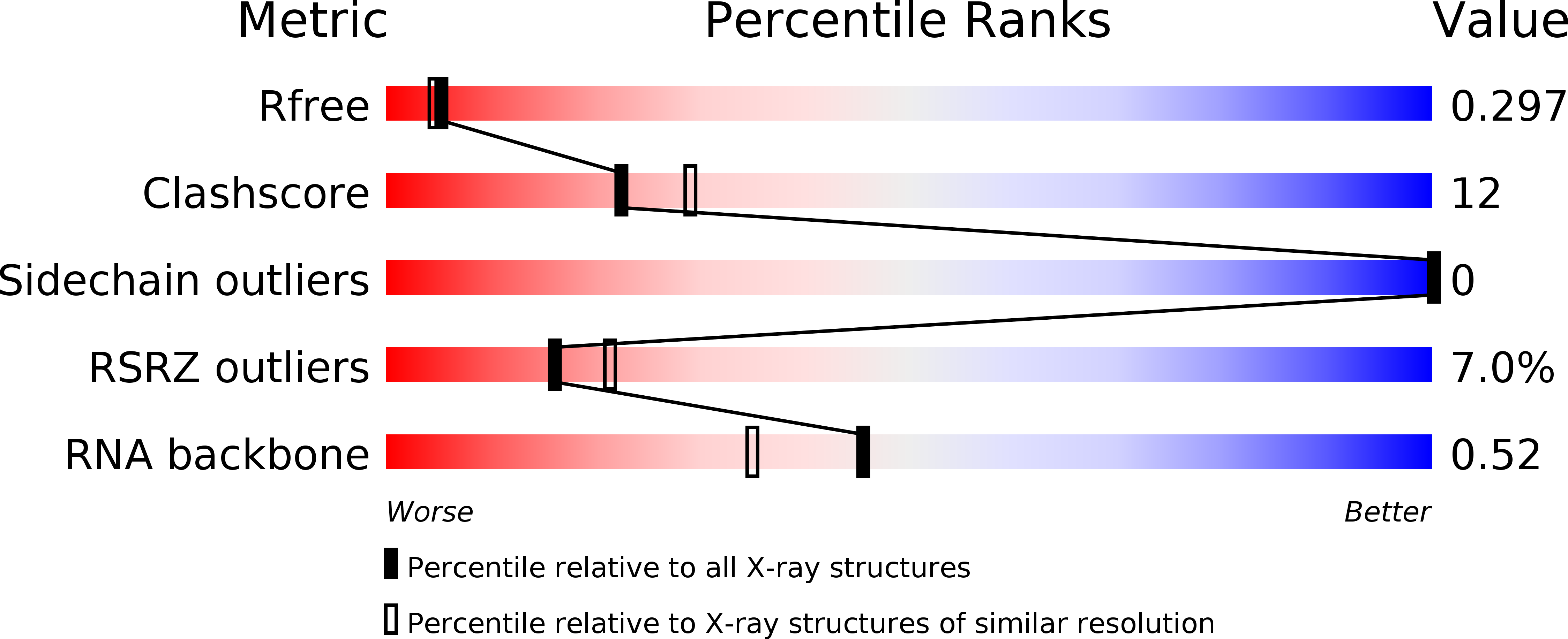
    6QN3

  • PubMed Abstract: 

    We have determined the structure of the glutamine-II riboswitch ligand binding domain using X-ray crystallography. The structure was solved using a novel combination of homology modeling and molecular replacement. The structure comprises three coaxial helical domains, the central one of which is a pseudoknot with partial triplex character. The major groove of this helix provides the binding site for L-glutamine, which is extensively hydrogen bonded to the RNA. Atomic mutation of the RNA at the ligand binding site leads to loss of binding shown by isothermal titration calorimetry, explaining the specificity of the riboswitch. A metal ion also plays an important role in ligand binding. This is directly bonded to a glutamine carboxylate oxygen atom, and its remaining inner-sphere water molecules make hydrogen bonding interactions with the RNA.


  • Organizational Affiliation

    Cancer Research UK Nucleic Acid Structure Research Group, MSI/WTB Complex, The University of Dundee, Dow Street, Dundee DD1 5EH, UK.


Macromolecules
Find similar nucleic acids by:  (by identity cutoff)  |  3D Structure
Entity ID: 1
MoleculeChains LengthOrganismImage
RNA (51-MER)
A, B
50Prochlorococcus sp.
Sequence Annotations
Expand
  • Reference Sequence
Small Molecules
Ligands 3 Unique
IDChains Name / Formula / InChI Key2D Diagram3D Interactions
GLN
Query on GLN

Download Ideal Coordinates CCD File 
G [auth A],
L [auth B]
GLUTAMINE
C5 H10 N2 O3
ZDXPYRJPNDTMRX-VKHMYHEASA-N
BR
Query on BR

Download Ideal Coordinates CCD File 
D [auth A],
E [auth A],
J [auth B],
K [auth B]
BROMIDE ION
Br
CPELXLSAUQHCOX-UHFFFAOYSA-M
MG
Query on MG

Download Ideal Coordinates CCD File 
C [auth A],
F [auth A],
H [auth B],
I [auth B]
MAGNESIUM ION
Mg
JLVVSXFLKOJNIY-UHFFFAOYSA-N
Experimental Data & Validation

Experimental Data

  • Method: X-RAY DIFFRACTION
  • Resolution: 2.30 Å
  • R-Value Free: 0.294 
  • R-Value Work: 0.242 
  • R-Value Observed: 0.245 
  • Space Group: C 2 2 21
Unit Cell:
Length ( Å )Angle ( ˚ )
a = 76.502α = 90
b = 115.725β = 90
c = 80.582γ = 90
Software Package:
Software NamePurpose
PHENIXrefinement
xia2data reduction
xia2data scaling
PHASERphasing

Structure Validation

View Full Validation Report


Entry History & Funding Information

Deposition Data


Funding OrganizationLocationGrant Number
Cancer Research UKUnited KingdomA18604

Revision History  (Full details and data files)

  • Version 1.0: 2019-06-12
    Type: Initial release
  • Version 1.1: 2019-07-03
    Changes: Data collection, Database references
  • Version 1.2: 2019-08-21
    Changes: Data collection
  • Version 1.3: 2019-08-28
    Changes: Data collection, Database references