6QFA

CryoEM structure of a beta3K279T GABA(A)R homomer in complex with histamine and megabody Mb25


Experimental Data Snapshot

  • Method: ELECTRON MICROSCOPY
  • Resolution: 2.49 Å
  • Aggregation State: PARTICLE 
  • Reconstruction Method: SINGLE PARTICLE 

wwPDB Validation   3D Report Full Report


This is version 1.0 of the entry. See complete history


Literature

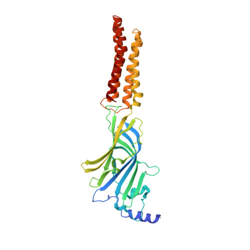
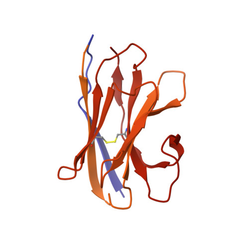
Megabodies expand the nanobody toolkit for protein structure determination by single-particle cryo-EM.

Uchanski, T.Masiulis, S.Fischer, B.Kalichuk, V.Lopez-Sanchez, U.Zarkadas, E.Weckener, M.Sente, A.Ward, P.Wohlkonig, A.Zogg, T.Remaut, H.Naismith, J.H.Nury, H.Vranken, W.Aricescu, A.R.Pardon, E.Steyaert, J.

(2021) Nat Methods 18: 60-68

  • DOI: https://doi.org/10.1038/s41592-020-01001-6
  • Primary Citation of Related Structures:  
    6QFA, 6XUX, 6XV8, 6XVI

  • PubMed Abstract: 

    Nanobodies are popular and versatile tools for structural biology. They have a compact single immunoglobulin domain organization, bind target proteins with high affinities while reducing their conformational heterogeneity and stabilize multi-protein complexes. Here we demonstrate that engineered nanobodies can also help overcome two major obstacles that limit the resolution of single-particle cryo-electron microscopy reconstructions: particle size and preferential orientation at the water-air interfaces. We have developed and characterized constructs, termed megabodies, by grafting nanobodies onto selected protein scaffolds to increase their molecular weight while retaining the full antigen-binding specificity and affinity. We show that the megabody design principles are applicable to different scaffold proteins and recognition domains of compatible geometries and are amenable for efficient selection from yeast display libraries. Moreover, we demonstrate that megabodies can be used to obtain three-dimensional reconstructions for membrane proteins that suffer from severe preferential orientation or are otherwise too small to allow accurate particle alignment.


  • Organizational Affiliation

    Structural Biology Brussels, Vrije Universiteit Brussel, VUB, Brussels, Belgium.


Macromolecules
Find similar proteins by:  (by identity cutoff)  |  3D Structure
Entity ID: 1
MoleculeChains Sequence LengthOrganismDetailsImage
Gamma-aminobutyric acid receptor subunit beta-3,Gamma-aminobutyric acid receptor subunit beta-3A [auth B],
G [auth C],
H [auth D],
I [auth E],
J [auth A]
341Homo sapiensMutation(s): 1 
Gene Names: GABRB3
Membrane Entity: Yes 
UniProt & NIH Common Fund Data Resources
Find proteins for P28472 (Homo sapiens)
Explore P28472 
Go to UniProtKB:  P28472
PHAROS:  P28472
GTEx:  ENSG00000166206 
Entity Groups  
Sequence Clusters30% Identity50% Identity70% Identity90% Identity95% Identity100% Identity
UniProt GroupP28472
Sequence Annotations
Expand
  • Reference Sequence
Find similar proteins by:  (by identity cutoff)  |  3D Structure
Entity ID: 2
MoleculeChains Sequence LengthOrganismDetailsImage
Outer membrane protein,Outer membrane protein,Outer membrane protein,Outer membrane protein,Uncharacterized protein,Uncharacterized protein,Mb-c7HopQ-Nb25B [auth K],
C [auth O],
D [auth N],
E [auth M],
F [auth L]
522Helicobacter pylori G27Lama glamaMutation(s): 0 
Gene Names: hopQHPG27_1120
Membrane Entity: Yes 
UniProt
Find proteins for B5Z8H1 (Helicobacter pylori (strain G27))
Explore B5Z8H1 
Go to UniProtKB:  B5Z8H1
Entity Groups  
Sequence Clusters30% Identity50% Identity70% Identity90% Identity95% Identity100% Identity
UniProt GroupB5Z8H1
Sequence Annotations
Expand
  • Reference Sequence
Oligosaccharides

Help

Entity ID: 3
MoleculeChains Length2D Diagram Glycosylation3D Interactions
alpha-D-mannopyranose-(1-3)-[alpha-D-mannopyranose-(1-6)]beta-D-mannopyranose-(1-4)-2-acetamido-2-deoxy-beta-D-glucopyranose-(1-4)-2-acetamido-2-deoxy-beta-D-glucopyranoseK [auth F],
L [auth G],
M [auth H],
N [auth I],
O [auth J]
5N/AN-Glycosylation
Small Molecules
Ligands 2 Unique
IDChains Name / Formula / InChI Key2D Diagram3D Interactions
NAG
Query on NAG

Download Ideal Coordinates CCD File 
P [auth B],
R [auth C],
T [auth D],
V [auth E],
X [auth A]
2-acetamido-2-deoxy-beta-D-glucopyranose
C8 H15 N O6
OVRNDRQMDRJTHS-FMDGEEDCSA-N
HSM (Subject of Investigation/LOI)
Query on HSM

Download Ideal Coordinates CCD File 
Q [auth B],
S [auth C],
U [auth D],
W [auth E],
Y [auth A]
HISTAMINE
C5 H9 N3
NTYJJOPFIAHURM-UHFFFAOYSA-N
Experimental Data & Validation

Experimental Data

  • Method: ELECTRON MICROSCOPY
  • Resolution: 2.49 Å
  • Aggregation State: PARTICLE 
  • Reconstruction Method: SINGLE PARTICLE 
EM Software:
TaskSoftware PackageVersion
MODEL REFINEMENTPHENIX
RECONSTRUCTIONRELION3.0

Structure Validation

View Full Validation Report


Entry History & Funding Information

Deposition Data


Funding OrganizationLocationGrant Number
Medical Research Council (United Kingdom)United KingdomMC_UP_1201/15
Medical Research Council (United Kingdom)United KingdomL009609/1
European UnionBelgium731005
Grenoble Instruct-ERIC CenterBelgium--
Research Foundation - FlandersBelgium--

Revision History  (Full details and data files)

  • Version 1.0: 2021-08-04
    Type: Initial release