6PX5

CRYSTAL STRUCTURE OF HUMAN MEIZOTHROMBIN DESF1 MUTANT S195A bound with PPACK


Experimental Data Snapshot

  • Method: X-RAY DIFFRACTION
  • Resolution: 2.40 Å
  • R-Value Free: 0.282 
  • R-Value Work: 0.223 
  • R-Value Observed: 0.226 

wwPDB Validation   3D Report Full Report


Ligand Structure Quality Assessment 


This is version 2.1 of the entry. See complete history


Literature

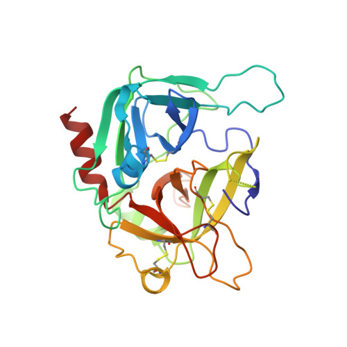
Residues W215, E217 and E192 control the allosteric E*-E equilibrium of thrombin.

Pelc, L.A.Koester, S.K.Chen, Z.Gistover, N.E.Di Cera, E.

(2019) Sci Rep 9: 12304-12304

  • DOI: https://doi.org/10.1038/s41598-019-48839-1
  • Primary Citation of Related Structures:  
    6P9U, 6PX5

  • PubMed Abstract: 

    A pre-existing, allosteric equilibrium between closed (E*) and open (E) conformations of the active site influences the level of activity in the trypsin fold and defines ligand binding according to the mechanism of conformational selection. Using the clotting protease thrombin as a model system, we investigate the molecular determinants of the E*-E equilibrium through rapid kinetics and X-ray structural biology. The equilibrium is controlled by three residues positioned around the active site. W215 on the 215-217 segment defining the west wall of the active site controls the rate of transition from E to E* through hydrophobic interaction with F227. E192 on the opposite 190-193 segment defining the east wall of the active site controls the rate of transition from E* to E through electrostatic repulsion of E217. The side chain of E217 acts as a lever that moves the entire 215-217 segment in the E*-E equilibrium. Removal of this side chain converts binding to the active site to a simple lock-and-key mechanism and freezes the conformation in a state intermediate between E* and E. These findings reveal a simple framework to understand the molecular basis of a key allosteric property of the trypsin fold.


  • Organizational Affiliation

    Edward A. Doisy Department of Biochemistry and Molecular Biology, Saint Louis University School of Medicine, St. Louis, MO, 63104, USA.


Macromolecules
Find similar proteins by:  (by identity cutoff)  |  3D Structure
Entity ID: 1
MoleculeChains Sequence LengthOrganismDetailsImage
ProthrombinA [auth X]152Homo sapiensMutation(s): 0 
Gene Names: F2
EC: 3.4.21.5
UniProt & NIH Common Fund Data Resources
Find proteins for P00734 (Homo sapiens)
Explore P00734 
Go to UniProtKB:  P00734
PHAROS:  P00734
GTEx:  ENSG00000180210 
Entity Groups  
Sequence Clusters30% Identity50% Identity70% Identity90% Identity95% Identity100% Identity
UniProt GroupP00734
Sequence Annotations
Expand
  • Reference Sequence
Find similar proteins by:  (by identity cutoff)  |  3D Structure
Entity ID: 2
MoleculeChains Sequence LengthOrganismDetailsImage
Prothrombin259Homo sapiensMutation(s): 1 
Gene Names: F2
EC: 3.4.21.5
UniProt & NIH Common Fund Data Resources
Find proteins for P00734 (Homo sapiens)
Explore P00734 
Go to UniProtKB:  P00734
PHAROS:  P00734
GTEx:  ENSG00000180210 
Entity Groups  
Sequence Clusters30% Identity50% Identity70% Identity90% Identity95% Identity100% Identity
UniProt GroupP00734
Sequence Annotations
Expand
  • Reference Sequence
Oligosaccharides

Help

Entity ID: 3
MoleculeChains Length2D Diagram Glycosylation3D Interactions
2-acetamido-2-deoxy-beta-D-glucopyranose-(1-4)-2-acetamido-2-deoxy-beta-D-glucopyranoseC [auth A]2N-Glycosylation
Glycosylation Resources
GlyTouCan:  G42666HT
GlyCosmos:  G42666HT
GlyGen:  G42666HT
Biologically Interesting Molecules (External Reference) 1 Unique
Experimental Data & Validation

Experimental Data

  • Method: X-RAY DIFFRACTION
  • Resolution: 2.40 Å
  • R-Value Free: 0.282 
  • R-Value Work: 0.223 
  • R-Value Observed: 0.226 
  • Space Group: P 32 2 1
Unit Cell:
Length ( Å )Angle ( ˚ )
a = 100.134α = 90
b = 100.134β = 90
c = 118.679γ = 120
Software Package:
Software NamePurpose
REFMACrefinement
PDB_EXTRACTdata extraction
HKL-2000data reduction
HKL-2000data scaling
MOLREPphasing

Structure Validation

View Full Validation Report


Ligand Structure Quality Assessment 


Entry History & Funding Information

Deposition Data


Funding OrganizationLocationGrant Number
National Institutes of Health/National Institute of Dental and Craniofacial Research (NIH/NIDCR)United States--

Revision History  (Full details and data files)

  • Version 1.0: 2019-09-04
    Type: Initial release
  • Version 1.1: 2019-12-11
    Changes: Author supporting evidence
  • Version 2.0: 2020-07-29
    Type: Remediation
    Reason: Carbohydrate remediation
    Changes: Atomic model, Data collection, Derived calculations, Structure summary
  • Version 2.1: 2023-10-11
    Changes: Data collection, Database references, Refinement description, Structure summary