


The Crystal Structure of the Manganese Superoxide Dismutase from Geobacillus stearothermophilus: Parker and Blake (1988) Revisited
Adams, J.J., Morton, C.J., Parker, M.W.(2020) Aust J Chem 73: 145-150
Experimental Data Snapshot
wwPDB Validation 3D Report Full Report

(2020) Aust J Chem 73: 145-150
Entity ID: 1 | |||||
|---|---|---|---|---|---|
| Molecule | Chains | Sequence Length | Organism | Details | Image |
| Superoxide dismutase | 202 | Geobacillus stearothermophilus | Mutation(s): 0 EC: 1.15.1.1 | 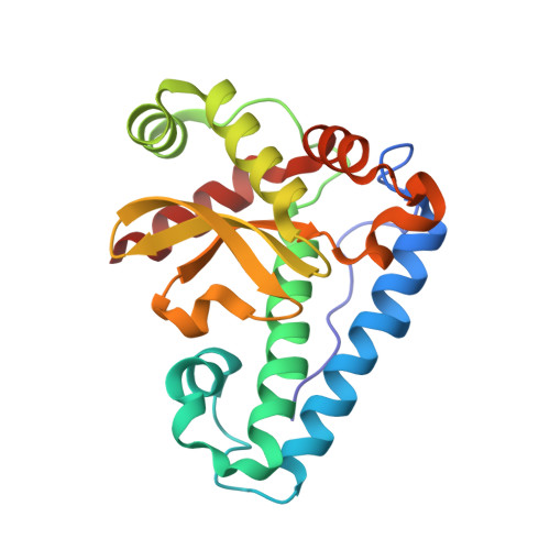 | |
UniProt | |||||
Find proteins for P00449 (Geobacillus stearothermophilus) Explore P00449 Go to UniProtKB: P00449 | |||||
Entity Groups | |||||
| Sequence Clusters | 30% Identity50% Identity70% Identity90% Identity95% Identity100% Identity | ||||
| UniProt Group | P00449 | ||||
Sequence AnnotationsExpand | |||||
| |||||
| Ligands 1 Unique | |||||
|---|---|---|---|---|---|
| ID | Chains | Name / Formula / InChI Key | 2D Diagram | 3D Interactions | |
| MN Query on MN | C [auth A], D [auth B] | MANGANESE (II) ION Mn WAEMQWOKJMHJLA-UHFFFAOYSA-N |  | ||
| Length ( Å ) | Angle ( ˚ ) |
|---|---|
| a = 72.2 | α = 90 |
| b = 111.1 | β = 90 |
| c = 51.1 | γ = 90 |
| Software Name | Purpose |
|---|---|
| PHENIX | refinement |
| PDB_EXTRACT | data extraction |
| MOSFLM | data reduction |
| in-house | data scaling |
| in-house | phasing |
RCSB PDB (citation) is hosted by
RCSB PDB is a member of the

