6P79

Engineered single chain antibody C9+C14 ScFv


Experimental Data Snapshot

  • Method: X-RAY DIFFRACTION
  • Resolution: 1.58 Å
  • R-Value Free: 0.202 
  • R-Value Work: 0.174 
  • R-Value Observed: 0.176 

wwPDB Validation   3D Report Full Report


This is version 1.2 of the entry. See complete history


Literature

Computer-based Engineering of Thermostabilized Antibody Fragments.

Lee, J.Der, B.S.Karamitros, C.S.Li, W.Marshall, N.M.Lungu, O.I.Miklos, A.E.Xu, J.Kang, T.H.Lee, C.H.Tan, B.Hughes, R.A.Jung, S.T.Ippolito, G.C.Gray, J.J.Zhang, Y.Kuhlman, B.Georgiou, G.Ellington, A.D.

(2020) AIChE J 66

  • DOI: https://doi.org/10.1002/aic.16864
  • Primary Citation of Related Structures:  
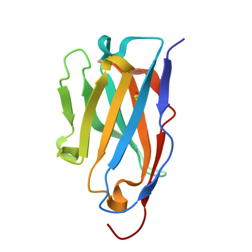
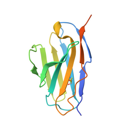
    6P79

  • PubMed Abstract: 

    We used the molecular modeling program Rosetta to identify clusters of amino acid substitutions in antibody fragments (scFvs and scAbs) that improve global protein stability and resistance to thermal deactivation. Using this methodology, we increased the melting temperature (T m ) and resistance to heat treatment of an antibody fragment that binds to the Clostridium botulinum hemagglutinin protein (anti-HA33). Two designed antibody fragment variants with two amino acid replacement clusters, designed to stabilize local regions, were shown to have both higher T m compared to the parental scFv and importantly, to retain full antigen binding activity after 2 hours of incubation at 70 °C. The crystal structure of one thermostabilized scFv variants was solved at 1.6 Å and shown to be in close agreement with the RosettaAntibody model prediction.


  • Organizational Affiliation

    Thayer School of Engineering, Dartmouth College, Hanover, NH 03755.


Macromolecules
Find similar proteins by:  (by identity cutoff)  |  3D Structure
Entity ID: 1
MoleculeChains Sequence LengthOrganismDetailsImage
Engineered antibody heavy chainA [auth H]120Mus musculusMutation(s): 0 
Entity Groups  
Sequence Clusters30% Identity50% Identity70% Identity90% Identity95% Identity100% Identity
Sequence Annotations
Expand
  • Reference Sequence
Find similar proteins by:  (by identity cutoff)  |  3D Structure
Entity ID: 2
MoleculeChains Sequence LengthOrganismDetailsImage
Engineered antibody light chainB [auth L]127Mus musculusMutation(s): 0 
Entity Groups  
Sequence Clusters30% Identity50% Identity70% Identity90% Identity95% Identity100% Identity
Sequence Annotations
Expand
  • Reference Sequence
Experimental Data & Validation

Experimental Data

  • Method: X-RAY DIFFRACTION
  • Resolution: 1.58 Å
  • R-Value Free: 0.202 
  • R-Value Work: 0.174 
  • R-Value Observed: 0.176 
  • Space Group: P 21 21 21
Unit Cell:
Length ( Å )Angle ( ˚ )
a = 44.282α = 90
b = 66.696β = 90
c = 74.955γ = 90
Software Package:
Software NamePurpose
PHENIXrefinement
HKL-3000data reduction
HKL-3000data scaling
PHENIXphasing

Structure Validation

View Full Validation Report


Entry History 

Deposition Data

Revision History  (Full details and data files)

  • Version 1.0: 2020-04-15
    Type: Initial release
  • Version 1.1: 2020-05-13
    Changes: Database references
  • Version 1.2: 2023-10-11
    Changes: Data collection, Database references, Refinement description