6P58

Dark and Steady State-Illuminated Crystal Structure of Cyanobacteriochrome Receptor PixJ at 150K


Experimental Data Snapshot

  • Method: X-RAY DIFFRACTION
  • Resolution: 1.50 Å
  • R-Value Free: 0.196 
  • R-Value Work: 0.159 
  • R-Value Observed: 0.161 

wwPDB Validation   3D Report Full Report

Currently 6P58 does not have a validation slider image.


This is version 1.3 of the entry. See complete history


Literature

Photoreversible interconversion of a phytochrome photosensory module in the crystalline state.

Burgie, E.S.Clinger, J.A.Miller, M.D.Brewster, A.S.Aller, P.Butryn, A.Fuller, F.D.Gul, S.Young, I.D.Pham, C.C.Kim, I.S.Bhowmick, A.O'Riordan, L.J.Sutherlin, K.D.Heinemann, J.V.Batyuk, A.Alonso-Mori, R.Hunter, M.S.Koglin, J.E.Yano, J.Yachandra, V.K.Sauter, N.K.Cohen, A.E.Kern, J.Orville, A.M.Phillips Jr., G.N.Vierstra, R.D.

(2020) Proc Natl Acad Sci U S A 117: 300-307

  • DOI: https://doi.org/10.1073/pnas.1912041116
  • Primary Citation of Related Structures:  
    6P58, 6PRU, 6PRY, 6UPP

  • PubMed Abstract: 

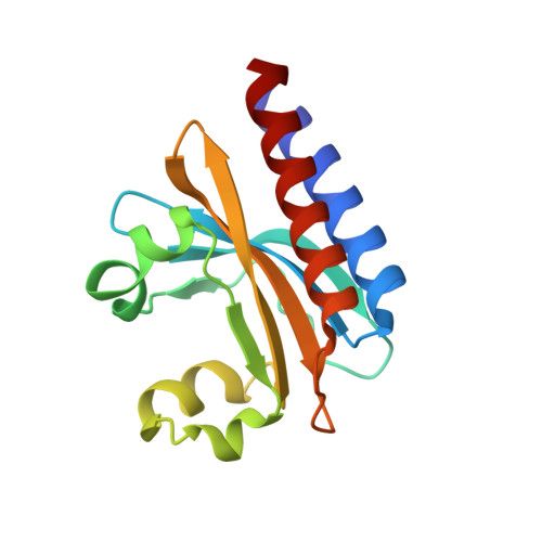
    A major barrier to defining the structural intermediates that arise during the reversible photointerconversion of phytochromes between their biologically inactive and active states has been the lack of crystals that faithfully undergo this transition within the crystal lattice. Here, we describe a crystalline form of the cyclic GMP phosphodiesterases/adenylyl cyclase/FhlA (GAF) domain from the cyanobacteriochrome PixJ in Thermosynechococcus elongatus assembled with phycocyanobilin that permits reversible photoconversion between the blue light-absorbing Pb and green light-absorbing Pg states, as well as thermal reversion of Pg back to Pb. The X-ray crystallographic structure of Pb matches previous models, including autocatalytic conversion of phycocyanobilin to phycoviolobilin upon binding and its tandem thioether linkage to the GAF domain. Cryocrystallography at 150 K, which compared diffraction data from a single crystal as Pb or after irradiation with blue light, detected photoconversion product(s) based on F obs - F obs difference maps that were consistent with rotation of the bonds connecting pyrrole rings C and D. Further spectroscopic analyses showed that phycoviolobilin is susceptible to X-ray radiation damage, especially as Pg, during single-crystal X-ray diffraction analyses, which could complicate fine mapping of the various intermediate states. Fortunately, we found that PixJ crystals are amenable to serial femtosecond crystallography (SFX) analyses using X-ray free-electron lasers (XFELs). As proof of principle, we solved by room temperature SFX the GAF domain structure of Pb to 1.55-Å resolution, which was strongly congruent with synchrotron-based models. Analysis of these crystals by SFX should now enable structural characterization of the early events that drive phytochrome photoconversion.


  • Organizational Affiliation

    Department of Biology, Washington University in St. Louis, St. Louis, MO 63130.


Macromolecules
Find similar proteins by:  (by identity cutoff)  |  3D Structure
Entity ID: 1
MoleculeChains Sequence LengthOrganismDetailsImage
Methyl-accepting chemotaxis protein
A, B
150Thermosynechococcus vestitus BP-1Mutation(s): 1 
Gene Names: tll0569
UniProt
Find proteins for Q8DLC7 (Thermosynechococcus vestitus (strain NIES-2133 / IAM M-273 / BP-1))
Explore Q8DLC7 
Go to UniProtKB:  Q8DLC7
Entity Groups  
Sequence Clusters30% Identity50% Identity70% Identity90% Identity95% Identity100% Identity
UniProt GroupQ8DLC7
Sequence Annotations
Expand
  • Reference Sequence
Small Molecules
Ligands 3 Unique
IDChains Name / Formula / InChI Key2D Diagram3D Interactions
VRB (Subject of Investigation/LOI)
Query on VRB

Download Ideal Coordinates CCD File 
C [auth A],
F [auth B]
Phycoviolobilin, blue light-absorbing form
C33 H42 N4 O6
KTSIPBCNIBDFRK-JTNIOKRPSA-N
EDO
Query on EDO

Download Ideal Coordinates CCD File 
G [auth B],
J [auth B]
1,2-ETHANEDIOL
C2 H6 O2
LYCAIKOWRPUZTN-UHFFFAOYSA-N
MG
Query on MG

Download Ideal Coordinates CCD File 
D [auth A],
E [auth A],
H [auth B],
I [auth B]
MAGNESIUM ION
Mg
JLVVSXFLKOJNIY-UHFFFAOYSA-N
Experimental Data & Validation

Experimental Data

  • Method: X-RAY DIFFRACTION
  • Resolution: 1.50 Å
  • R-Value Free: 0.196 
  • R-Value Work: 0.159 
  • R-Value Observed: 0.161 
  • Space Group: P 21 21 21
Unit Cell:
Length ( Å )Angle ( ˚ )
a = 41.618α = 90
b = 61.052β = 90
c = 116.148γ = 90
Software Package:
Software NamePurpose
Aimlessdata scaling
PHENIXrefinement
PDB_EXTRACTdata extraction
DIALSdata reduction
PHENIXphasing

Structure Validation

View Full Validation Report

Currently 6P58 does not have a validation slider image.


Entry History & Funding Information

Deposition Data


Funding OrganizationLocationGrant Number
National Science Foundation (NSF, United States)United StatesSTC-1231306
National Institutes of Health/National Institute of General Medical Sciences (NIH/NIGMS)United StatesGM127892

Revision History  (Full details and data files)

  • Version 1.0: 2019-12-18
    Type: Initial release
  • Version 1.1: 2020-01-01
    Changes: Database references
  • Version 1.2: 2020-01-22
    Changes: Database references
  • Version 1.3: 2023-10-11
    Changes: Data collection, Database references, Derived calculations, Refinement description, Structure summary