6P38

Crystal Structure Analysis of TAF1 Bromodomain


Experimental Data Snapshot

  • Method: X-RAY DIFFRACTION
  • Resolution: 2.80 Å
  • R-Value Free: 0.219 
  • R-Value Work: 0.206 
  • R-Value Observed: 0.206 

wwPDB Validation   3D Report Full Report


Ligand Structure Quality Assessment 


This is version 1.2 of the entry. See complete history


Literature

Dual Inhibition of TAF1 and BET Bromodomains from the BI-2536 Kinase Inhibitor Scaffold.

Remillard, D.Buckley, D.L.Seo, H.S.Ferguson, F.M.Dhe-Paganon, S.Bradner, J.E.Gray, N.S.

(2019) ACS Med Chem Lett 10: 1443-1449

  • DOI: https://doi.org/10.1021/acsmedchemlett.9b00243
  • Primary Citation of Related Structures:  
    6P38

  • PubMed Abstract: 

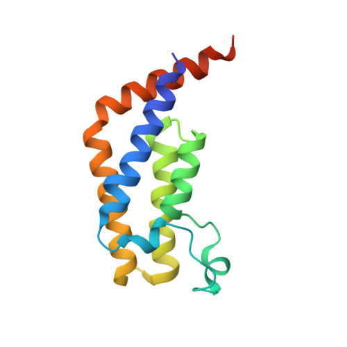
    Recent reports have highlighted the dual bromodomains of TAF1 (TAF1(1,2)) as synergistic with BET inhibition in cellular cancer models, engendering interest in TAF/BET polypharmacology. Here, we examine structure activity relationships within the BI-2536 PLK1 kinase inhibitor scaffold, previously reported to bind BRD4. We examine binding by this ligand to TAF1(2) and apply structure guided design strategies to discriminate binding to both the PLK1 kinase and BRD4(1) bromodomain while retaining activity on TAF1(2). Through this effort we discover potent dual inhibitors of TAF1(2)/BRD4(1), as well as biased derivatives showing marked TAF1 selectivity. We resolve X-ray crystallographic data sets to examine the mechanisms of the observed TAF1 selectivity and to provide a resource for further development of this scaffold.


  • Organizational Affiliation

    Department of Cancer Biology, Dana Farber Cancer Institute, Boston Massachusetts 02115, United States.


Macromolecules
Find similar proteins by:  (by identity cutoff)  |  3D Structure
Entity ID: 1
MoleculeChains Sequence LengthOrganismDetailsImage
Transcription initiation factor TFIID subunit 1137Homo sapiensMutation(s): 0 
Gene Names: TAF1BA2RCCG1CCGSTAF2A
EC: 2.3.1.48 (PDB Primary Data), 2.7.11.1 (PDB Primary Data)
UniProt & NIH Common Fund Data Resources
Find proteins for P21675 (Homo sapiens)
Explore P21675 
Go to UniProtKB:  P21675
PHAROS:  P21675
GTEx:  ENSG00000147133 
Entity Groups  
Sequence Clusters30% Identity50% Identity70% Identity90% Identity95% Identity100% Identity
UniProt GroupP21675
Sequence Annotations
Expand
  • Reference Sequence
Small Molecules
Ligands 1 Unique
IDChains Name / Formula / InChI Key2D Diagram3D Interactions
NQV (Subject of Investigation/LOI)
Query on NQV

Download Ideal Coordinates CCD File 
B [auth A]4-{[(3R)-4-cyclopentyl-1,3-dimethyl-2-oxo-1,2,3,4-tetrahydropyrido[2,3-b]pyrazin-6-yl]amino}-N-(1-methylpiperidin-4-yl)-3-[(propan-2-yl)oxy]benzamide
C30 H42 N6 O3
AGCMTUNUCHFYSQ-HXUWFJFHSA-N
Experimental Data & Validation

Experimental Data

  • Method: X-RAY DIFFRACTION
  • Resolution: 2.80 Å
  • R-Value Free: 0.219 
  • R-Value Work: 0.206 
  • R-Value Observed: 0.206 
  • Space Group: P 31 2 1
Unit Cell:
Length ( Å )Angle ( ˚ )
a = 95.89α = 90
b = 95.89β = 90
c = 94.22γ = 120
Software Package:
Software NamePurpose
XDSdata reduction
xia2data scaling
PHENIXrefinement
PDB_EXTRACTdata extraction
PHASERphasing

Structure Validation

View Full Validation Report


Ligand Structure Quality Assessment 


Entry History & Funding Information

Deposition Data


Funding OrganizationLocationGrant Number
National Institutes of Health/National Cancer Institute (NIH/NCI)United States--

Revision History  (Full details and data files)

  • Version 1.0: 2020-05-27
    Type: Initial release
  • Version 1.1: 2020-12-09
    Changes: Database references
  • Version 1.2: 2023-10-11
    Changes: Data collection, Database references, Refinement description