6OT4

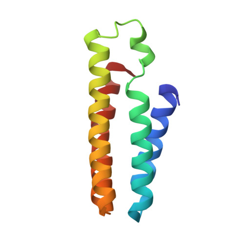
Bimetallic dodecameric cage design 2 (BMC2) from cytochrome cb562


Experimental Data Snapshot

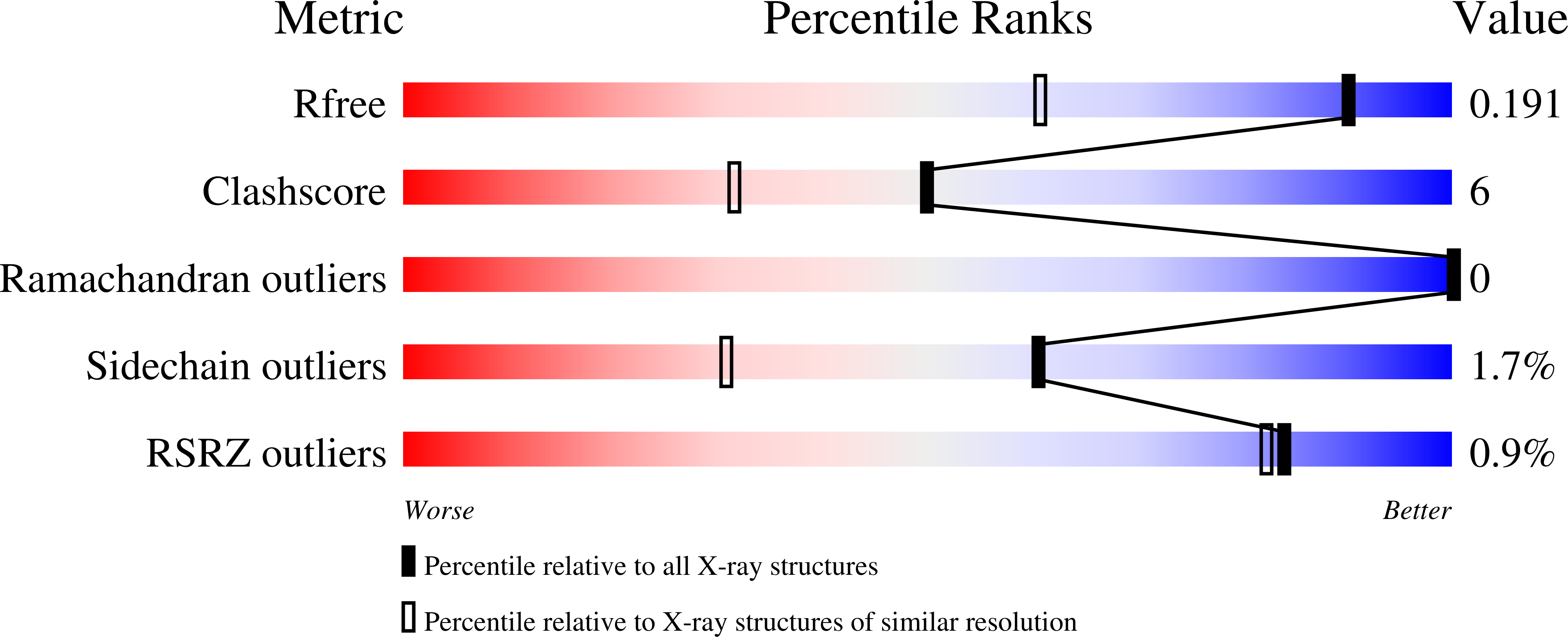
  • Method: X-RAY DIFFRACTION
  • Resolution: 1.40 Å
  • R-Value Free: 0.191 
  • R-Value Work: 0.166 
  • R-Value Observed: 0.166 

wwPDB Validation   3D Report Full Report


Ligand Structure Quality Assessment 


This is version 1.3 of the entry. See complete history


Literature

Constructing protein polyhedra via orthogonal chemical interactions.

Golub, E.Subramanian, R.H.Esselborn, J.Alberstein, R.G.Bailey, J.B.Chiong, J.A.Yan, X.Booth, T.Baker, T.S.Tezcan, F.A.

(2020) Nature 578: 172-176

  • DOI: https://doi.org/10.1038/s41586-019-1928-2
  • Primary Citation of Related Structures:  
    6OT4, 6OT7, 6OT8, 6OT9, 6OVH

  • PubMed Abstract: 

    Many proteins exist naturally as symmetrical homooligomers or homopolymers 1 . The emergent structural and functional properties of such protein assemblies have inspired extensive efforts in biomolecular design 2-5 . As synthesized by ribosomes, proteins are inherently asymmetric. Thus, they must acquire multiple surface patches that selectively associate to generate the different symmetry elements needed to form higher-order architectures 1,6 -a daunting task for protein design. Here we address this problem using an inorganic chemical approach, whereby multiple modes of protein-protein interactions and symmetry are simultaneously achieved by selective, 'one-pot' coordination of soft and hard metal ions. We show that a monomeric protein (protomer) appropriately modified with biologically inspired hydroxamate groups and zinc-binding motifs assembles through concurrent Fe 3+ and Zn 2+ coordination into discrete dodecameric and hexameric cages. Our cages closely resemble natural polyhedral protein architectures 7,8 and are, to our knowledge, unique among designed systems 9-13 in that they possess tightly packed shells devoid of large apertures. At the same time, they can assemble and disassemble in response to diverse stimuli, owing to their heterobimetallic construction on minimal interprotein-bonding footprints. With stoichiometries ranging from [2 Fe:9 Zn:6 protomers] to [8 Fe:21 Zn:12 protomers], these protein cages represent some of the compositionally most complex protein assemblies-or inorganic coordination complexes-obtained by design.


  • Organizational Affiliation

    Department of Chemistry and Biochemistry, University of California, San Diego, La Jolla, CA, USA.


Macromolecules
Find similar proteins by:  (by identity cutoff)  |  3D Structure
Entity ID: 1
MoleculeChains Sequence LengthOrganismDetailsImage
Soluble cytochrome b562
A, B, C, D
106Escherichia coliMutation(s): 20 
Gene Names: cybC
UniProt
Find proteins for P0ABE7 (Escherichia coli)
Explore P0ABE7 
Go to UniProtKB:  P0ABE7
Entity Groups  
Sequence Clusters30% Identity50% Identity70% Identity90% Identity95% Identity100% Identity
UniProt GroupP0ABE7
Sequence Annotations
Expand
  • Reference Sequence
Small Molecules
Ligands 4 Unique
IDChains Name / Formula / InChI Key2D Diagram3D Interactions
HEC
Query on HEC

Download Ideal Coordinates CCD File 
E [auth A],
L [auth B],
R [auth C],
W [auth D]
HEME C
C34 H34 Fe N4 O4
HXQIYSLZKNYNMH-LJNAALQVSA-N
HAE (Subject of Investigation/LOI)
Query on HAE

Download Ideal Coordinates CCD File 
F [auth A]
G [auth A]
M [auth B]
N [auth B]
S [auth C]
F [auth A],
G [auth A],
M [auth B],
N [auth B],
S [auth C],
T [auth C],
X [auth D],
Y [auth D]
ACETOHYDROXAMIC ACID
C2 H5 N O2
RRUDCFGSUDOHDG-UHFFFAOYSA-N
ZN (Subject of Investigation/LOI)
Query on ZN

Download Ideal Coordinates CCD File 
I [auth A]
J [auth A]
K [auth A]
P [auth B]
U [auth C]
I [auth A],
J [auth A],
K [auth A],
P [auth B],
U [auth C],
V [auth C]
ZINC ION
Zn
PTFCDOFLOPIGGS-UHFFFAOYSA-N
FE (Subject of Investigation/LOI)
Query on FE

Download Ideal Coordinates CCD File 
H [auth A],
O [auth B],
Q [auth B],
Z [auth D]
FE (III) ION
Fe
VTLYFUHAOXGGBS-UHFFFAOYSA-N
Experimental Data & Validation

Experimental Data

  • Method: X-RAY DIFFRACTION
  • Resolution: 1.40 Å
  • R-Value Free: 0.191 
  • R-Value Work: 0.166 
  • R-Value Observed: 0.166 
  • Space Group: H 3 2
Unit Cell:
Length ( Å )Angle ( ˚ )
a = 126.14α = 90
b = 126.14β = 90
c = 168.21γ = 120
Software Package:
Software NamePurpose
XDSdata reduction
XSCALEdata scaling
PHENIXrefinement
PDB_EXTRACTdata extraction
PHASERphasing

Structure Validation

View Full Validation Report


Ligand Structure Quality Assessment 


Entry History & Funding Information

Deposition Data


Funding OrganizationLocationGrant Number
National Science Foundation (NSF, United States)United StatesDMR-1602537
Department of Energy (DOE, United States)United StatesDE-SC0003844
European Molecular Biology Organization (EMBO)European UnionALTF 1336-2015
German Research Foundation (DFG)Germany393131496

Revision History  (Full details and data files)

  • Version 1.0: 2020-01-29
    Type: Initial release
  • Version 1.1: 2020-02-05
    Changes: Database references
  • Version 1.2: 2020-02-19
    Changes: Database references
  • Version 1.3: 2023-10-11
    Changes: Data collection, Database references, Derived calculations, Refinement description