Allosteric regulation of menaquinone (vitamin K2) biosynthesis in the human pathogenMycobacterium tuberculosis.
Bashiri, G., Nigon, L.V., Jirgis, E.N.M., Ho, N.A.T., Stanborough, T., Dawes, S.S., Baker, E.N., Bulloch, E.M.M., Johnston, J.M.(2020) J Biol Chem 295: 3759-3770
- PubMed: 32029475
- DOI: https://doi.org/10.1074/jbc.RA119.012158
- Primary Citation of Related Structures:
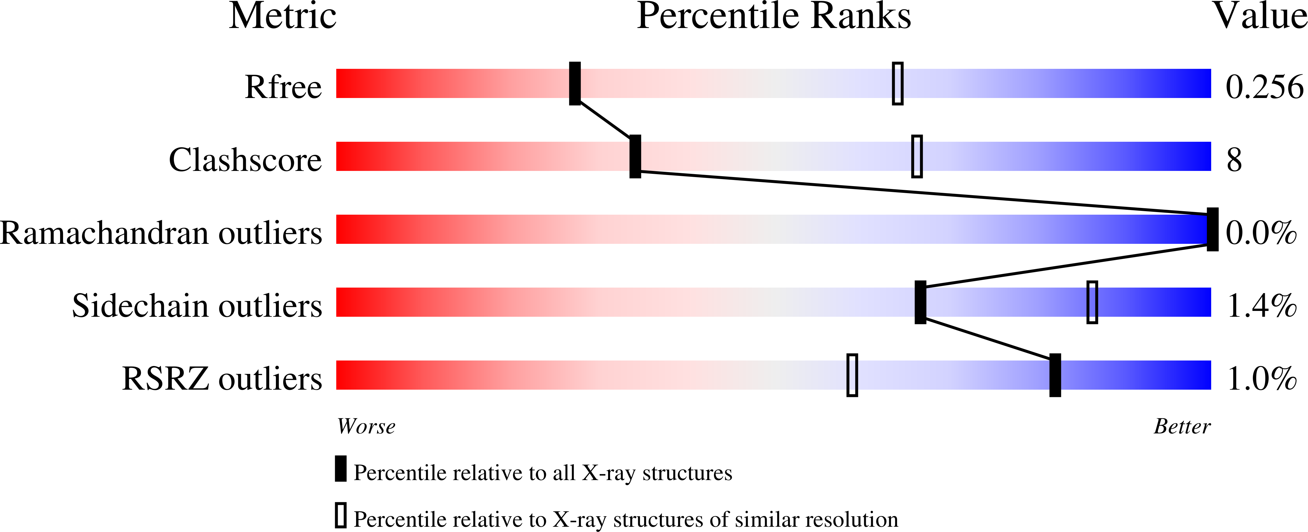
6O04, 6O0G, 6O0J, 6O0N - PubMed Abstract:
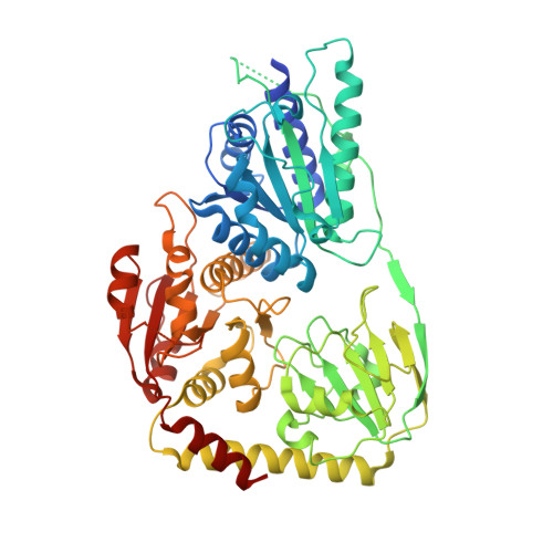
Menaquinone (vitamin K 2 ) plays a vital role in energy generation and environmental adaptation in many bacteria, including the human pathogen Mycobacterium tuberculosis ( Mtb ). Although menaquinone levels are known to be tightly linked to the cellular redox/energy status of the cell, the regulatory mechanisms underpinning this phenomenon are unclear. The first committed step in menaquinone biosynthesis is catalyzed by MenD, a thiamine diphosphate-dependent enzyme comprising three domains. Domains I and III form the MenD active site, but no function has yet been ascribed to domain II. Here, we show that the last cytosolic metabolite in the menaquinone biosynthesis pathway, 1,4-dihydroxy-2-naphthoic acid (DHNA), binds to domain II of Mtb -MenD and inhibits its activity. Using X-ray crystallography of four apo- and cofactor-bound Mtb -MenD structures, along with several spectroscopy assays, we identified three arginine residues (Arg-97, Arg-277, and Arg-303) that are important for both enzyme activity and the feedback inhibition by DHNA. Among these residues, Arg-277 appeared to be particularly important for signal propagation from the allosteric site to the active site. This is the first evidence of feedback regulation of the menaquinone biosynthesis pathway in bacteria, identifying a protein-level regulatory mechanism that controls menaquinone levels within the cell and may therefore represent a good target for disrupting menaquinone biosynthesis in M. tuberculosis .
Organizational Affiliation:
Laboratory of Structural Biology, School of Biological Sciences and Maurice Wilkins Centre for Molecular Biodiscovery, University of Auckland, Private Bag 92019, Auckland 1010, New Zealand.