6NPW

SSu72/Sympk in complex with Ser2/Ser5 phosphorylated peptide


Experimental Data Snapshot

  • Method: X-RAY DIFFRACTION
  • Resolution: 2.49 Å
  • R-Value Free: 0.235 
  • R-Value Work: 0.189 
  • R-Value Observed: 0.191 

wwPDB Validation   3D Report Full Report


This is version 1.2 of the entry. See complete history


Literature

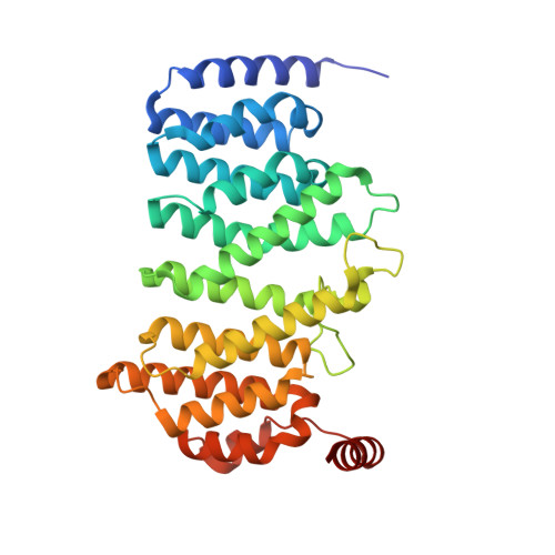
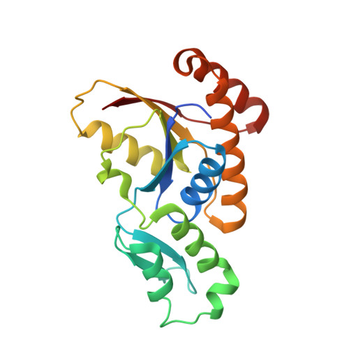
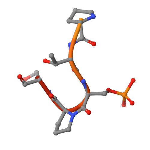
Structural determinants for accurate dephosphorylation of RNA polymerase II by its cognate C-terminal domain (CTD) phosphatase during eukaryotic transcription.

Irani, S.Sipe, S.N.Yang, W.Burkholder, N.T.Lin, B.Sim, K.Matthews, W.L.Brodbelt, J.S.Zhang, Y.

(2019) J Biol Chem 294: 8592-8605

  • DOI: https://doi.org/10.1074/jbc.RA119.007697
  • Primary Citation of Related Structures:  
    6NPW

  • PubMed Abstract: 

    The C-terminal domain (CTD) of RNA polymerase II contains a repetitive heptad sequence (YSPTSPS) whose phosphorylation states coordinate eukaryotic transcription by recruiting protein regulators. The precise placement and removal of phosphate groups on specific residues of the CTD are critical for the fidelity and effectiveness of RNA polymerase II-mediated transcription. During transcriptional elongation, phosphoryl-Ser 5 (pSer 5 ) is gradually dephosphorylated by CTD phosphatases, whereas Ser 2 phosphorylation accumulates. Using MS, X-ray crystallography, protein engineering, and immunoblotting analyses, here we investigated the structure and function of SSU72 homolog, RNA polymerase II CTD phosphatase (Ssu72, from Drosophila melanogaster ), an essential CTD phosphatase that dephosphorylates pSer 5 at the transition from elongation to termination, to determine the mechanism by which Ssu72 distinguishes the highly similar pSer 2 and pSer 5 CTDs. We found that Ssu72 dephosphorylates pSer 5 effectively but only has low activities toward pSer 7 and pSer 2 The structural analysis revealed that Ssu72 requires that the proline residue in the substrate's SP motif is in the cis configuration, forming a tight β-turn for recognition by Ssu72. We also noted that residues flanking the SP motif, such as the bulky Tyr 1 next to Ser 2 , prevent the formation of such configuration and enable Ssu72 to distinguish among the different SP motifs. The phosphorylation of Tyr 1 further prohibited Ssu72 binding to pSer 2 and thereby prevented untimely Ser 2 dephosphorylation. Our results reveal critical roles for Tyr 1 in differentiating the phosphorylation states of Ser 2 /Ser 5 of CTD in RNA polymerase II that occur at different stages of transcription.


  • Organizational Affiliation

    Department of Chemical Engineering, The University of Texas, Austin, Texas 78712; Department of Molecular Biosciences, The University of Texas, Austin, Texas 78712.


Macromolecules
Find similar proteins by:  (by identity cutoff)  |  3D Structure
Entity ID: 1
MoleculeChains Sequence LengthOrganismDetailsImage
Symplekin
A, C
339Drosophila melanogasterMutation(s): 0 
Gene Names: SymCG2097
UniProt
Find proteins for Q8MSU4 (Drosophila melanogaster)
Explore Q8MSU4 
Go to UniProtKB:  Q8MSU4
Entity Groups  
Sequence Clusters30% Identity50% Identity70% Identity90% Identity95% Identity100% Identity
UniProt GroupQ8MSU4
Sequence Annotations
Expand
  • Reference Sequence
Find similar proteins by:  (by identity cutoff)  |  3D Structure
Entity ID: 2
MoleculeChains Sequence LengthOrganismDetailsImage
Ssu72 ortholog, LD40846p
B, D
200Drosophila melanogasterMutation(s): 2 
Gene Names: Ssu72CG14216Dmel_CG14216
EC: 3.1.3 (PDB Primary Data), 3.1.3.16 (PDB Primary Data), 3.1.3.41 (PDB Primary Data)
UniProt
Find proteins for Q9VWE4 (Drosophila melanogaster)
Explore Q9VWE4 
Go to UniProtKB:  Q9VWE4
Entity Groups  
Sequence Clusters30% Identity50% Identity70% Identity90% Identity95% Identity100% Identity
UniProt GroupQ9VWE4
Sequence Annotations
Expand
  • Reference Sequence

Find similar proteins by:  Sequence   |   3D Structure  

Entity ID: 3
MoleculeChains Sequence LengthOrganismDetailsImage
Ser2/Ser5 phosphorylated peptide19Saccharomyces cerevisiae RM11-1aMutation(s): 0 
UniProt
Find proteins for P04050 (Saccharomyces cerevisiae (strain ATCC 204508 / S288c))
Explore P04050 
Go to UniProtKB:  P04050
Entity Groups  
Sequence Clusters30% Identity50% Identity70% Identity90% Identity95% Identity100% Identity
UniProt GroupP04050
Sequence Annotations
Expand
  • Reference Sequence
Small Molecules
Ligands 1 Unique
IDChains Name / Formula / InChI Key2D Diagram3D Interactions
PO4
Query on PO4

Download Ideal Coordinates CCD File 
F [auth B]PHOSPHATE ION
O4 P
NBIIXXVUZAFLBC-UHFFFAOYSA-K
Modified Residues  1 Unique
IDChains TypeFormula2D DiagramParent
SEP
Query on SEP
E
L-PEPTIDE LINKINGC3 H8 N O6 PSER
Experimental Data & Validation

Experimental Data

  • Method: X-RAY DIFFRACTION
  • Resolution: 2.49 Å
  • R-Value Free: 0.235 
  • R-Value Work: 0.189 
  • R-Value Observed: 0.191 
  • Space Group: P 4
Unit Cell:
Length ( Å )Angle ( ˚ )
a = 127.702α = 90
b = 127.702β = 90
c = 105.979γ = 90
Software Package:
Software NamePurpose
PHENIXrefinement
HKL-2000data reduction
HKL-2000data scaling
PHASERphasing

Structure Validation

View Full Validation Report


Entry History 

Deposition Data

Revision History  (Full details and data files)

  • Version 1.0: 2019-04-24
    Type: Initial release
  • Version 1.1: 2019-06-05
    Changes: Data collection, Database references
  • Version 1.2: 2023-10-11
    Changes: Data collection, Database references, Refinement description