6NJU

Mouse endonuclease G mutant H97A bound to A-DNA


Experimental Data Snapshot

  • Method: X-RAY DIFFRACTION
  • Resolution: 2.35 Å
  • R-Value Free: 0.291 
  • R-Value Work: 0.222 
  • R-Value Observed: 0.226 

wwPDB Validation   3D Report Full Report

Currently 6NJU does not have a validation slider image.


This is version 1.5 of the entry. See complete history


Literature

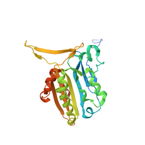
Structural adaptation of vertebrate endonuclease G for 5-hydroxymethylcytosine recognition and function.

Vander Zanden, C.M.Czarny, R.S.Ho, E.N.Robertson, A.B.Ho, P.S.

(2020) Nucleic Acids Res 48: 3962-3974

  • DOI: https://doi.org/10.1093/nar/gkaa117
  • Primary Citation of Related Structures:  
    6NJT, 6NJU

  • PubMed Abstract: 

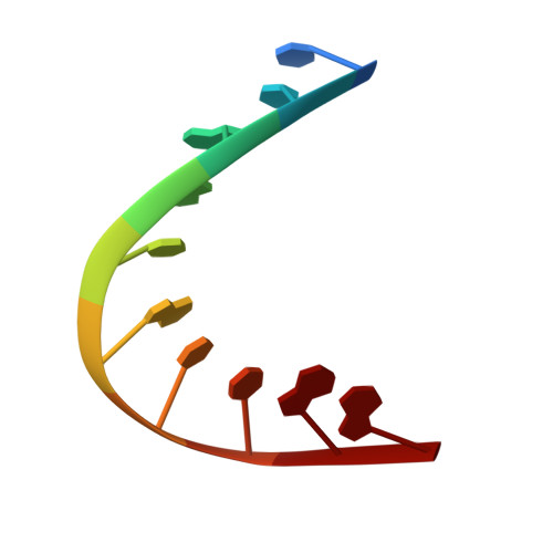
    Modified DNA bases functionally distinguish the taxonomic forms of life-5-methylcytosine separates prokaryotes from eukaryotes and 5-hydroxymethylcytosine (5hmC) invertebrates from vertebrates. We demonstrate here that mouse endonuclease G (mEndoG) shows specificity for both 5hmC and Holliday junctions. The enzyme has higher affinity (>50-fold) for junctions over duplex DNAs. A 5hmC-modification shifts the position of the cut site and increases the rate of DNA cleavage in modified versus unmodified junctions. The crystal structure of mEndoG shows that a cysteine (Cys69) is positioned to recognize 5hmC through a thiol-hydroxyl hydrogen bond. Although this Cys is conserved from worms to mammals, a two amino acid deletion in the vertebrate relative to the invertebrate sequence unwinds an α-helix, placing the thiol of Cys69 into the mEndoG active site. Mutations of Cys69 with alanine or serine show 5hmC-specificity that mirrors the hydrogen bonding potential of the side chain (C-H < S-H < O-H). A second orthogonal DNA binding site identified in the mEndoG structure accommodates a second arm of a junction. Thus, the specificity of mEndoG for 5hmC and junctions derives from structural adaptations that distinguish the vertebrate from the invertebrate enzyme, thereby thereby supporting a role for 5hmC in recombination processes.


  • Organizational Affiliation

    Department of Biochemistry & Molecular Biology, Colorado State University, Fort Collins, CO 80523-1870, USA.


Macromolecules

Find similar proteins by:  (by identity cutoff)  |  3D Structure
Entity ID: 1
MoleculeChains Sequence LengthOrganismDetailsImage
Endonuclease G, mitochondrial
A, B, C, D
253Mus musculusMutation(s): 1 
Gene Names: Endog
EC: 3.1.30
UniProt & NIH Common Fund Data Resources
Find proteins for O08600 (Mus musculus)
Explore O08600 
Go to UniProtKB:  O08600
IMPC:  MGI:1261433
Entity Groups  
Sequence Clusters30% Identity50% Identity70% Identity90% Identity95% Identity100% Identity
UniProt GroupO08600
Sequence Annotations
Expand
  • Reference Sequence

Find similar nucleic acids by:  Sequence   |   3D Structure  

Entity ID: 2
MoleculeChains LengthOrganismImage
DNA (5'-D(CCGGCGCCGG)-3')10Mus musculus
Sequence Annotations
Expand
  • Reference Sequence
Small Molecules
Ligands 3 Unique
IDChains Name / Formula / InChI Key2D Diagram3D Interactions
TRS
Query on TRS

Download Ideal Coordinates CCD File 
N [auth E]2-AMINO-2-HYDROXYMETHYL-PROPANE-1,3-DIOL
C4 H12 N O3
LENZDBCJOHFCAS-UHFFFAOYSA-O
CL
Query on CL

Download Ideal Coordinates CCD File 
G [auth A],
J [auth C],
K [auth C],
M [auth D]
CHLORIDE ION
Cl
VEXZGXHMUGYJMC-UHFFFAOYSA-M
MG
Query on MG

Download Ideal Coordinates CCD File 
F [auth A],
H [auth B],
I [auth C],
L [auth D]
MAGNESIUM ION
Mg
JLVVSXFLKOJNIY-UHFFFAOYSA-N
Experimental Data & Validation

Experimental Data

  • Method: X-RAY DIFFRACTION
  • Resolution: 2.35 Å
  • R-Value Free: 0.291 
  • R-Value Work: 0.222 
  • R-Value Observed: 0.226 
  • Space Group: P 65 2 2
Unit Cell:
Length ( Å )Angle ( ˚ )
a = 107.983α = 90
b = 107.983β = 90
c = 357.419γ = 120
Software Package:
Software NamePurpose
PHENIXrefinement
XDSdata reduction
PHASERphasing
Cootmodel building
Aimlessdata scaling

Structure Validation

View Full Validation Report

Currently 6NJU does not have a validation slider image.


Entry History & Funding Information

Deposition Data


Funding OrganizationLocationGrant Number
National Science Foundation (NSF, United States)United States1515521

Revision History  (Full details and data files)

  • Version 1.0: 2020-01-08
    Type: Initial release
  • Version 1.1: 2020-01-15
    Changes: Derived calculations
  • Version 1.2: 2020-01-29
    Changes: Derived calculations
  • Version 1.3: 2020-03-11
    Changes: Database references
  • Version 1.4: 2020-04-22
    Changes: Database references
  • Version 1.5: 2023-10-11
    Changes: Data collection, Database references, Derived calculations, Refinement description