6MV4

CRYSTAL STRUCTURE OF HUMAN COAGULATION FACTOR IXa


Experimental Data Snapshot

  • Method: X-RAY DIFFRACTION
  • Resolution: 1.37 Å
  • R-Value Free: 0.210 
  • R-Value Work: 0.177 
  • R-Value Observed: 0.178 

wwPDB Validation   3D Report Full Report

Currently 6MV4 does not have a validation slider image.


This is version 1.3 of the entry. See complete history


Literature

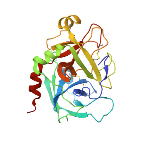
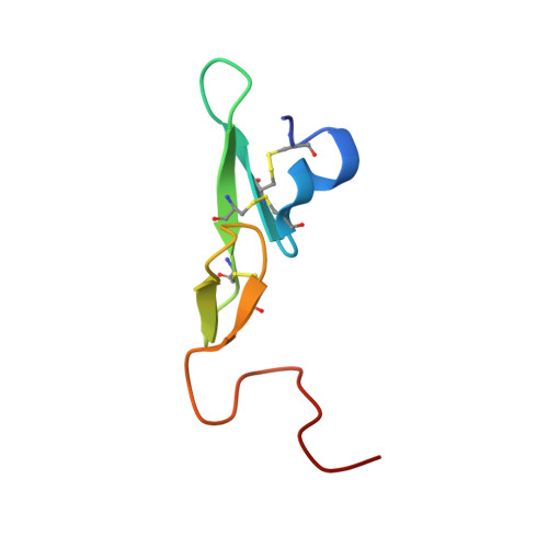
Sodium-site in serine protease domain of human coagulation factor IXa: evidence from the crystal structure and molecular dynamics simulations study.

Vadivel, K.Schreuder, H.A.Liesum, A.Schmidt, A.E.Goldsmith, G.Bajaj, S.P.

(2019) J Thromb Haemost 17: 574-584

  • DOI: https://doi.org/10.1111/jth.14401
  • Primary Citation of Related Structures:  
    6MV4

  • PubMed Abstract: 

    Essentials Consensus sequence and biochemical data suggest a Na + -site in the factor (F) IXa protease domain. X-ray structure of the FIXa EGF2/protease domain at 1.37 Å reveals a Na + -site not observed earlier. Molecular dynamics simulations data support that Na +  ± Ca 2+ promote FIXa protease domain stability. Sulfate ions found in the protease domain mimic heparin sulfate binding mode in FIXa. SUMMARY: Background Activated coagulation factor IX (FIXa) consists of a γ-carboxyglutamic acid domain, two epidermal growth factor-like (EGF) domains, and a C-terminal protease domain. Consensus sequence and biochemical data support the existence of a Na + -site in the FIXa protease domain. However, soaking experiments or crystals grown in high concentration of ammonium sulfate did not reveal a Na + -site in wild-type or mutant FIXa EGF2/protease domain structure. Objective Determine the structure of the FIXa EGF2/protease domain in the presence of Na + ; perform molecular dynamics (MD) simulations to explore the role of Na + in stabilizing FIXa structure. Methods Crystallography, MD simulations, and modeling heparin binding to FIXa. Results Crystal structure at 1.37-Å resolution revealed that Na + is coordinated to carbonyl groups of residues 184A, 185, 221A, and 224 in the FIXa protease domain. The Na + -site in FIXa is similar to that of FXa and is linked to the Asp189 S1-site. In MD simulations, Na + reduced fluctuations in residues 217-225 (Na + -loop) and 70-80 (Ca 2+ -loop), whereas Ca 2+ reduced fluctuations only in residues of the Ca 2+ -loop. Ca 2+ and Na + together reduced fluctuations in residues of the Ca 2+ -loop and Na + -loop (residues 70-80, 183-194, and 217-225). Moreover, we observed four sulfate ions that make salt bridges with FIXa protease domain Arg/Lys residues, which have been implicated in heparin binding. Based upon locations of the sulfate ions, we modeled heparin binding to FIXa, which is similar to the heparin binding in thrombin. Conclusions The FIXa Na + -site in association with Ca 2+ contributes to stabilization of the FIXa protease domain. The heparin binding mode in FIXa is similar to that in thrombin.


  • Organizational Affiliation

    Department of Orthopaedic Surgery, University of California, Los Angeles, CA, USA.


Macromolecules
Find similar proteins by:  (by identity cutoff)  |  3D Structure
Entity ID: 1
MoleculeChains Sequence LengthOrganismDetailsImage
Coagulation factor IXA [auth H]235Homo sapiensMutation(s): 0 
Gene Names: F9
EC: 3.4.21.22
UniProt & NIH Common Fund Data Resources
Find proteins for P00740 (Homo sapiens)
Explore P00740 
Go to UniProtKB:  P00740
PHAROS:  P00740
GTEx:  ENSG00000101981 
Entity Groups  
Sequence Clusters30% Identity50% Identity70% Identity90% Identity95% Identity100% Identity
UniProt GroupP00740
Sequence Annotations
Expand
  • Reference Sequence
Find similar proteins by:  (by identity cutoff)  |  3D Structure
Entity ID: 2
MoleculeChains Sequence LengthOrganismDetailsImage
Coagulation factor IXB [auth L]54Homo sapiensMutation(s): 0 
Gene Names: F9
EC: 3.4.21.22
UniProt & NIH Common Fund Data Resources
Find proteins for P00740 (Homo sapiens)
Explore P00740 
Go to UniProtKB:  P00740
PHAROS:  P00740
GTEx:  ENSG00000101981 
Entity Groups  
Sequence Clusters30% Identity50% Identity70% Identity90% Identity95% Identity100% Identity
UniProt GroupP00740
Sequence Annotations
Expand
  • Reference Sequence
Small Molecules
Ligands 7 Unique
IDChains Name / Formula / InChI Key2D Diagram3D Interactions
PBZ
Query on PBZ

Download Ideal Coordinates CCD File 
L [auth H]P-AMINO BENZAMIDINE
C7 H10 N3
WPANETAWYGDRLL-UHFFFAOYSA-O
SO4
Query on SO4

Download Ideal Coordinates CCD File 
C [auth H]
D [auth H]
E [auth H]
F [auth H]
G [auth H]
C [auth H],
D [auth H],
E [auth H],
F [auth H],
G [auth H],
H
SULFATE ION
O4 S
QAOWNCQODCNURD-UHFFFAOYSA-L
EDO
Query on EDO

Download Ideal Coordinates CCD File 
I [auth H],
J [auth H]
1,2-ETHANEDIOL
C2 H6 O2
LYCAIKOWRPUZTN-UHFFFAOYSA-N
FMT
Query on FMT

Download Ideal Coordinates CCD File 
K [auth H]FORMIC ACID
C H2 O2
BDAGIHXWWSANSR-UHFFFAOYSA-N
CA
Query on CA

Download Ideal Coordinates CCD File 
M [auth H]CALCIUM ION
Ca
BHPQYMZQTOCNFJ-UHFFFAOYSA-N
CL
Query on CL

Download Ideal Coordinates CCD File 
O [auth H],
P [auth H],
Q [auth H]
CHLORIDE ION
Cl
VEXZGXHMUGYJMC-UHFFFAOYSA-M
NA
Query on NA

Download Ideal Coordinates CCD File 
N [auth H]SODIUM ION
Na
FKNQFGJONOIPTF-UHFFFAOYSA-N
Experimental Data & Validation

Experimental Data

  • Method: X-RAY DIFFRACTION
  • Resolution: 1.37 Å
  • R-Value Free: 0.210 
  • R-Value Work: 0.177 
  • R-Value Observed: 0.178 
  • Space Group: I 41 2 2
Unit Cell:
Length ( Å )Angle ( ˚ )
a = 109.758α = 90
b = 109.758β = 90
c = 112.458γ = 90
Software Package:
Software NamePurpose
BUSTERrefinement
XDSdata reduction
autoPROCdata scaling
Aimlessdata scaling
AMoREphasing

Structure Validation

View Full Validation Report

Currently 6MV4 does not have a validation slider image.


Entry History & Funding Information

Deposition Data


Funding OrganizationLocationGrant Number
National Institutes of Health/National Heart, Lung, and Blood Institute (NIH/NHLBI)United StatesR01HL141850

Revision History  (Full details and data files)

  • Version 1.0: 2019-02-20
    Type: Initial release
  • Version 1.1: 2019-04-10
    Changes: Data collection, Database references
  • Version 1.2: 2019-12-04
    Changes: Author supporting evidence
  • Version 1.3: 2023-10-11
    Changes: Data collection, Database references, Refinement description