6MS1

Crystal structure of the human Scribble PDZ1 domain bound to the PDZ-binding motif of APC


Experimental Data Snapshot

  • Method: X-RAY DIFFRACTION
  • Resolution: 1.35 Å
  • R-Value Free: 0.203 
  • R-Value Work: 0.178 
  • R-Value Observed: 0.179 

wwPDB Validation   3D Report Full Report

Currently 6MS1 does not have a validation slider image.


This is version 1.3 of the entry. See complete history


Literature

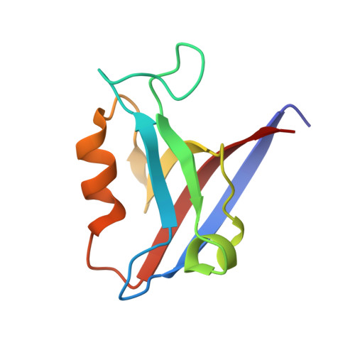
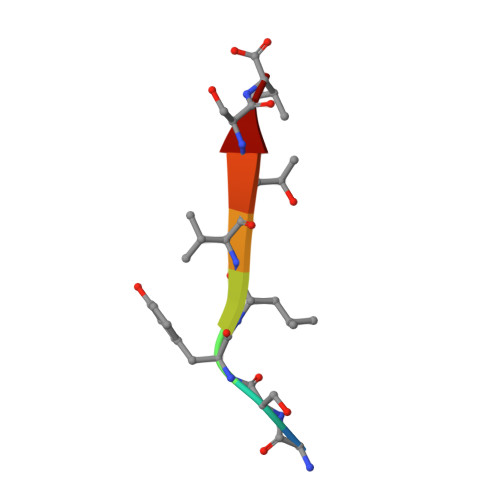
Crystal structure of the human Scribble PDZ1 domain bound to the PDZ-binding motif of APC.

How, J.Y.Caria, S.Humbert, P.O.Kvansakul, M.

(2019) FEBS Lett 593: 533-542

  • DOI: https://doi.org/10.1002/1873-3468.13329
  • Primary Citation of Related Structures:  
    6MS1

  • PubMed Abstract: 

    Scribble (SCRIB) is an important adaptor protein that controls the establishment and maintenance of apico-basal cell polarity. To better understand how SCRIB controls cell polarity signalling via its PDZ domains, we investigated human SCRIB interactions with adenomatous polyposis coli (APC). We show that SCRIB PDZ1, PDZ2 and PDZ3 are the major interactors with the APC PDZ-binding motif (PBM), whereas SCRIB PDZ4 does not show detectable binding to APC. We then determined the crystal structure of SCRIB PDZ1 domain bound to the APC PBM. Our findings reveal a previously unreported pattern of interactions between the SCRIB PDZ domain region with the C-terminal PDZ binding motif of APC, where SCRIB PDZ1 domain is the highest affinity site.


  • Organizational Affiliation

    Department of Biochemistry & Genetics, La Trobe Institute for Molecular Science, La Trobe University, Melbourne, Australia.


Macromolecules
Find similar proteins by:  (by identity cutoff)  |  3D Structure
Entity ID: 1
MoleculeChains Sequence LengthOrganismDetailsImage
Protein scribble homolog
A, B
94Homo sapiensMutation(s): 0 
Gene Names: SCRIBCRIB1KIAA0147LAP4SCRB1VARTUL
UniProt & NIH Common Fund Data Resources
Find proteins for Q14160 (Homo sapiens)
Explore Q14160 
Go to UniProtKB:  Q14160
PHAROS:  Q14160
GTEx:  ENSG00000180900 
Entity Groups  
Sequence Clusters30% Identity50% Identity70% Identity90% Identity95% Identity100% Identity
UniProt GroupQ14160
Sequence Annotations
Expand
  • Reference Sequence

Find similar proteins by:  Sequence   |   3D Structure  

Entity ID: 2
MoleculeChains Sequence LengthOrganismDetailsImage
APC C-terminus peptide
C, D
8Homo sapiensMutation(s): 0 
Sequence Annotations
Expand
  • Reference Sequence
Experimental Data & Validation

Experimental Data

  • Method: X-RAY DIFFRACTION
  • Resolution: 1.35 Å
  • R-Value Free: 0.203 
  • R-Value Work: 0.178 
  • R-Value Observed: 0.179 
  • Space Group: C 1 2 1
Unit Cell:
Length ( Å )Angle ( ˚ )
a = 62.338α = 90
b = 51.102β = 92.69
c = 57.051γ = 90
Software Package:
Software NamePurpose
PHENIXrefinement
Aimlessdata scaling
PHENIXphasing
xia2data reduction
Blu-Icedata collection
Cootmodel building

Structure Validation

View Full Validation Report

Currently 6MS1 does not have a validation slider image.


Entry History 

Deposition Data

Revision History  (Full details and data files)

  • Version 1.0: 2019-02-06
    Type: Initial release
  • Version 1.1: 2019-03-27
    Changes: Data collection, Database references
  • Version 1.2: 2020-01-08
    Changes: Author supporting evidence
  • Version 1.3: 2023-10-11
    Changes: Data collection, Database references, Refinement description