6MR1

RbcS-like subdomain of CcmM


Experimental Data Snapshot

  • Method: X-RAY DIFFRACTION
  • Resolution: 1.35 Å
  • R-Value Free: 0.195 
  • R-Value Work: 0.171 
  • R-Value Observed: 0.173 

wwPDB Validation   3D Report Full Report


This is version 1.5 of the entry. See complete history


Literature

The small RbcS-like domains of the beta-carboxysome structural protein CcmM bind RubisCO at a site distinct from that binding the RbcS subunit.

Ryan, P.Forrester, T.J.B.Wroblewski, C.Kenney, T.M.G.Kitova, E.N.Klassen, J.S.Kimber, M.S.

(2019) J Biol Chem 294: 2593-2603

  • DOI: https://doi.org/10.1074/jbc.RA118.006330
  • Primary Citation of Related Structures:  
    6MR1

  • PubMed Abstract: 

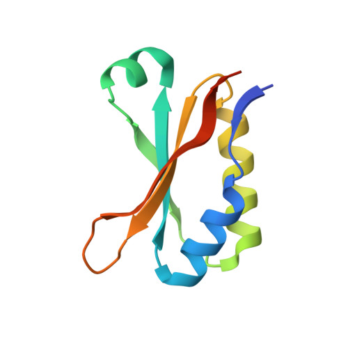
    Carboxysomes are compartments in bacterial cells that promote efficient carbon fixation by sequestering RubisCO and carbonic anhydrase within a protein shell that impedes CO 2 escape. The key to assembling this protein complex is CcmM, a multidomain protein whose C-terminal region is required for RubisCO recruitment. This CcmM region is built as a series of copies (generally 3-5) of a small domain, CcmM S , joined by unstructured linkers. CcmM S domains have weak, but significant, sequence identity to RubisCO's small subunit, RbcS, suggesting that CcmM binds RubisCO by displacing RbcS. We report here the 1.35-Å structure of the first Thermosynechococcus elongatus CcmM S domain, revealing that it adopts a compact, well-defined structure that resembles that of RbcS. CcmM S , however, lacked key RbcS RubisCO-binding determinants, most notably an extended N-terminal loop. Nevertheless, individual CcmM S domains are able to bind RubisCO in vitro with 1.16 μm affinity. Two or four linked CcmM S domains did not exhibit dramatic increases in this affinity, implying that short, disordered linkers may frustrate successive CcmM S domains attempting to simultaneously bind a single RubisCO oligomer. Size-exclusion chromatography-coupled right-angled light scattering (SEC-RALS) and native MS experiments indicated that multiple CcmM S domains can bind a single RubisCO holoenzyme and, moreover, that RbcS is not released from these complexes. CcmM S bound equally tightly to a RubisCO variant in which the α/β domain of RbcS was deleted, suggesting that CcmM S binds RubisCO independently of its RbcS subunit. We propose that, instead, the electropositive CcmM S may bind to an extended electronegative pocket between RbcL dimers.


  • Organizational Affiliation

    From the Department of Molecular and Cellular Biology, University of Guelph, Guelph, Ontario N1G 2W1, Canada and.


Macromolecules
Find similar proteins by:  (by identity cutoff)  |  3D Structure
Entity ID: 1
MoleculeChains Sequence LengthOrganismDetailsImage
Carbon dioxide concentrating mechanism protein
A, B
96Thermosynechococcus vestitus BP-1Mutation(s): 0 
Gene Names: ccmM
UniProt
Find proteins for Q8DKB5 (Thermosynechococcus vestitus (strain NIES-2133 / IAM M-273 / BP-1))
Explore Q8DKB5 
Go to UniProtKB:  Q8DKB5
Entity Groups  
Sequence Clusters30% Identity50% Identity70% Identity90% Identity95% Identity100% Identity
UniProt GroupQ8DKB5
Sequence Annotations
Expand
  • Reference Sequence
Small Molecules
Ligands 4 Unique
IDChains Name / Formula / InChI Key2D Diagram3D Interactions
SO4
Query on SO4

Download Ideal Coordinates CCD File 
D [auth A],
J [auth B]
SULFATE ION
O4 S
QAOWNCQODCNURD-UHFFFAOYSA-L
CO
Query on CO

Download Ideal Coordinates CCD File 
C [auth A],
F [auth B],
G [auth B]
COBALT (II) ION
Co
XLJKHNWPARRRJB-UHFFFAOYSA-N
SCN
Query on SCN

Download Ideal Coordinates CCD File 
H [auth B],
I [auth B]
THIOCYANATE ION
C N S
ZMZDMBWJUHKJPS-UHFFFAOYSA-M
CL
Query on CL

Download Ideal Coordinates CCD File 
E [auth A]CHLORIDE ION
Cl
VEXZGXHMUGYJMC-UHFFFAOYSA-M
Experimental Data & Validation

Experimental Data

  • Method: X-RAY DIFFRACTION
  • Resolution: 1.35 Å
  • R-Value Free: 0.195 
  • R-Value Work: 0.171 
  • R-Value Observed: 0.173 
  • Space Group: P 21 21 21
Unit Cell:
Length ( Å )Angle ( ˚ )
a = 28.9α = 90
b = 73.56β = 90
c = 76.57γ = 90
Software Package:
Software NamePurpose
PHENIXrefinement
XDSdata reduction
XSCALEdata scaling
PHENIXphasing

Structure Validation

View Full Validation Report


Entry History & Funding Information

Deposition Data


Funding OrganizationLocationGrant Number
Natural Sciences and Engineering Research Council (NSERC, Canada)Canada04045-2015

Revision History  (Full details and data files)

  • Version 1.0: 2019-01-02
    Type: Initial release
  • Version 1.1: 2019-01-09
    Changes: Data collection, Database references
  • Version 1.2: 2019-03-06
    Changes: Data collection, Database references
  • Version 1.3: 2019-11-20
    Changes: Derived calculations
  • Version 1.4: 2020-01-08
    Changes: Author supporting evidence
  • Version 1.5: 2024-03-13
    Changes: Data collection, Database references, Derived calculations