6MO6

Crystal structure of the selenomethionine-substituted human sulfide:quinone oxidoreductase


Experimental Data Snapshot

  • Method: X-RAY DIFFRACTION
  • Resolution: 2.59 Å
  • R-Value Free: 0.245 
  • R-Value Work: 0.187 
  • R-Value Observed: 0.190 

wwPDB Validation   3D Report Full Report


Ligand Structure Quality Assessment 


This is version 1.2 of the entry. See complete history


Literature

X-Ray Structure of Human Sulfide:Quinone Oxidoreductase: Insights into the Mechanism of Mitochondrial Hydrogen Sulfide Oxidation.

Jackson, M.R.Loll, P.J.Jorns, M.S.

(2019) Structure 27: 794-805.e4

  • DOI: https://doi.org/10.1016/j.str.2019.03.002
  • Primary Citation of Related Structures:  
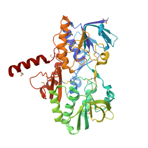
    6MO6, 6MP5

  • PubMed Abstract: 

    Hydrogen sulfide (H 2 S) is a gasotransmitter exhibiting pivotal functions in diverse biological processes, including activation of multiple cardioprotective pathways. Sulfide:quinone oxidoreductase (SQOR) is an integral membrane flavoprotein that catalyzes the first step in the mitochondrial metabolism of H 2 S. As such, it plays a critical role in controlling physiological levels of the gasotransmitter and has attracted keen interest as a potential drug target. We report the crystal structure of human SQOR, unraveling the molecular basis for the enzyme's ability to catalyze sulfane sulfur transfer reactions with structurally diverse acceptors. We demonstrate that human SQOR contains unique features: an electropositive surface depression implicated as a binding site for sulfane sulfur acceptors and postulated to funnel negatively charged substrates to a hydrophilic H 2 S-oxidizing active site, which is connected to a hydrophobic internal tunnel that binds coenzyme Q. These findings support a proposed model for catalysis and open the door for structure-based drug design.


  • Organizational Affiliation

    Biochemistry and Molecular Biology, Drexel University College of Medicine, 245 N. 15(th) Street, Philadelphia, PA 19102, USA.


Macromolecules
Find similar proteins by:  (by identity cutoff)  |  3D Structure
Entity ID: 1
MoleculeChains Sequence LengthOrganismDetailsImage
Sulfide:quinone oxidoreductase, mitochondrial
A, B, C, D
414Homo sapiensMutation(s): 0 
Gene Names: SQORSQRDLCGI-44
EC: 1.8.5
Membrane Entity: Yes 
UniProt & NIH Common Fund Data Resources
Find proteins for Q9Y6N5 (Homo sapiens)
Explore Q9Y6N5 
Go to UniProtKB:  Q9Y6N5
PHAROS:  Q9Y6N5
GTEx:  ENSG00000137767 
Entity Groups  
Sequence Clusters30% Identity50% Identity70% Identity90% Identity95% Identity100% Identity
UniProt GroupQ9Y6N5
Sequence Annotations
Expand
  • Reference Sequence
Small Molecules
Modified Residues  2 Unique
IDChains TypeFormula2D DiagramParent
CSS
Query on CSS
A, B, C, D
L-PEPTIDE LINKINGC3 H7 N O2 S2CYS
MSE
Query on MSE
A, B, C, D
L-PEPTIDE LINKINGC5 H11 N O2 SeMET
Experimental Data & Validation

Experimental Data

  • Method: X-RAY DIFFRACTION
  • Resolution: 2.59 Å
  • R-Value Free: 0.245 
  • R-Value Work: 0.187 
  • R-Value Observed: 0.190 
  • Space Group: P 61 2 2
Unit Cell:
Length ( Å )Angle ( ˚ )
a = 119.39α = 90
b = 119.39β = 90
c = 551.858γ = 120
Software Package:
Software NamePurpose
PHENIXrefinement
REFMACrefinement
MOSFLMdata reduction
Aimlessdata scaling
SHELXCDphasing

Structure Validation

View Full Validation Report


Ligand Structure Quality Assessment 


Entry History & Funding Information

Deposition Data


Funding OrganizationLocationGrant Number
National Institutes of Health/National Institute of General Medical Sciences (NIH/NIGMS)United StatesR01 GM107389
National Institutes of Health/National Heart, Lung, and Blood Institute (NIH/NHLBI)United StatesR41 HL134435

Revision History  (Full details and data files)

  • Version 1.0: 2019-04-10
    Type: Initial release
  • Version 1.1: 2019-05-15
    Changes: Data collection, Database references
  • Version 1.2: 2019-12-04
    Changes: Author supporting evidence