6ML1

Structure of the USP15 deubiquitinase domain in complex with an affinity-matured inhibitory Ubv


Experimental Data Snapshot

  • Method: X-RAY DIFFRACTION
  • Resolution: 1.90 Å
  • R-Value Free: 0.220 
  • R-Value Work: 0.189 
  • R-Value Observed: 0.191 

wwPDB Validation   3D Report Full Report


This is version 1.5 of the entry. See complete history


Literature

Structural and Functional Characterization of Ubiquitin Variant Inhibitors of USP15.

Teyra, J.Singer, A.U.Schmitges, F.W.Jaynes, P.Kit Leng Lui, S.Polyak, M.J.Fodil, N.Krieger, J.R.Tong, J.Schwerdtfeger, C.Brasher, B.B.Ceccarelli, D.F.J.Moffat, J.Sicheri, F.Moran, M.F.Gros, P.Eichhorn, P.J.A.Lenter, M.Boehmelt, G.Sidhu, S.S.

(2019) Structure 27: 590

  • DOI: https://doi.org/10.1016/j.str.2019.01.002
  • Primary Citation of Related Structures:  
    6CPM, 6CRN, 6DJ9, 6ML1

  • PubMed Abstract: 

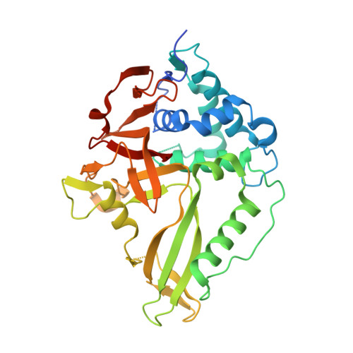
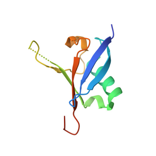
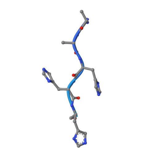
    The multi-domain deubiquitinase USP15 regulates diverse eukaryotic processes and has been implicated in numerous diseases. We developed ubiquitin variants (UbVs) that targeted either the catalytic domain or each of three adaptor domains in USP15, including the N-terminal DUSP domain. We also designed a linear dimer (diUbV), which targeted the DUSP and catalytic domains, and exhibited enhanced specificity and more potent inhibition of catalytic activity than either UbV alone. In cells, the UbVs inhibited the deubiquitination of two USP15 substrates, SMURF2 and TRIM25, and the diUbV inhibited the effects of USP15 on the transforming growth factor β pathway. Structural analyses revealed that three distinct UbVs bound to the catalytic domain and locked the active site in a closed, inactive conformation, and one UbV formed an unusual strand-swapped dimer and bound two DUSP domains simultaneously. These inhibitors will enable the study of USP15 function in oncology, neurology, immunology, and inflammation.


  • Organizational Affiliation

    The Donnelly Centre, University of Toronto, Toronto, ON M5S 3E1, Canada.


Macromolecules
Find similar proteins by:  (by identity cutoff)  |  3D Structure
Entity ID: 1
MoleculeChains Sequence LengthOrganismDetailsImage
Ubiquitin carboxyl-terminal hydrolase 15,Ubiquitin carboxyl-terminal hydrolase 15,Ubiquitin carboxyl-terminal hydrolase 15
A, B
349Homo sapiensMutation(s): 0 
Gene Names: USP15KIAA0529
EC: 3.4.19.12
UniProt & NIH Common Fund Data Resources
Find proteins for Q9Y4E8 (Homo sapiens)
Explore Q9Y4E8 
Go to UniProtKB:  Q9Y4E8
PHAROS:  Q9Y4E8
GTEx:  ENSG00000135655 
Entity Groups  
Sequence Clusters30% Identity50% Identity70% Identity90% Identity95% Identity100% Identity
UniProt GroupQ9Y4E8
Sequence Annotations
Expand
  • Reference Sequence
Find similar proteins by:  (by identity cutoff)  |  3D Structure
Entity ID: 2
MoleculeChains Sequence LengthOrganismDetailsImage
Ubiquitin variant 15.1aC [auth E],
D [auth C]
92Homo sapiensMutation(s): 0 
Gene Names: UBC
UniProt
Find proteins for A0A0L7RG06 (Habropoda laboriosa)
Explore A0A0L7RG06 
Go to UniProtKB:  A0A0L7RG06
Entity Groups  
Sequence Clusters30% Identity50% Identity70% Identity90% Identity95% Identity100% Identity
UniProt GroupA0A0L7RG06
Sequence Annotations
Expand
  • Reference Sequence
Find similar proteins by:  (by identity cutoff)  |  3D Structure
Entity ID: 3
MoleculeChains Sequence LengthOrganismDetailsImage
Proteolyzed N-terminal tag of Ubv.15.1a constructE [auth G]26Escherichia coliMutation(s): 0 
Entity Groups  
Sequence Clusters30% Identity50% Identity70% Identity90% Identity95% Identity100% Identity
Sequence Annotations
Expand
  • Reference Sequence
Small Molecules
Ligands 6 Unique
IDChains Name / Formula / InChI Key2D Diagram3D Interactions
MES
Query on MES

Download Ideal Coordinates CCD File 
R [auth C]2-(N-MORPHOLINO)-ETHANESULFONIC ACID
C6 H13 N O4 S
SXGZJKUKBWWHRA-UHFFFAOYSA-N
ZN
Query on ZN

Download Ideal Coordinates CCD File 
H [auth A],
L [auth B]
ZINC ION
Zn
PTFCDOFLOPIGGS-UHFFFAOYSA-N
EDO
Query on EDO

Download Ideal Coordinates CCD File 
M [auth B]1,2-ETHANEDIOL
C2 H6 O2
LYCAIKOWRPUZTN-UHFFFAOYSA-N
CA
Query on CA

Download Ideal Coordinates CCD File 
F [auth A],
G [auth A],
K [auth B],
Q [auth C]
CALCIUM ION
Ca
BHPQYMZQTOCNFJ-UHFFFAOYSA-N
CL
Query on CL

Download Ideal Coordinates CCD File 
N [auth B]CHLORIDE ION
Cl
VEXZGXHMUGYJMC-UHFFFAOYSA-M
NA
Query on NA

Download Ideal Coordinates CCD File 
I [auth A],
J [auth A],
O [auth B],
P [auth B]
SODIUM ION
Na
FKNQFGJONOIPTF-UHFFFAOYSA-N
Experimental Data & Validation

Experimental Data

  • Method: X-RAY DIFFRACTION
  • Resolution: 1.90 Å
  • R-Value Free: 0.220 
  • R-Value Work: 0.189 
  • R-Value Observed: 0.191 
  • Space Group: P 1 21 1
Unit Cell:
Length ( Å )Angle ( ˚ )
a = 42.77α = 90
b = 115.29β = 91.92
c = 95.32γ = 90
Software Package:
Software NamePurpose
PHENIXrefinement
MOSFLMdata reduction
SCALAdata scaling
PHASERphasing

Structure Validation

View Full Validation Report


Entry History 

Deposition Data

Revision History  (Full details and data files)

  • Version 1.0: 2019-01-23
    Type: Initial release
  • Version 1.1: 2019-02-13
    Changes: Data collection, Database references
  • Version 1.2: 2019-04-17
    Changes: Data collection, Database references
  • Version 1.3: 2023-02-08
    Changes: Advisory, Database references, Derived calculations
  • Version 1.4: 2023-10-04
    Changes: Data collection
  • Version 1.5: 2023-10-25
    Changes: Refinement description