6MAL

Structure of human Nocturnin C-terminal domain


Experimental Data Snapshot

  • Method: X-RAY DIFFRACTION
  • Resolution: 2.60 Å
  • R-Value Free: 0.307 
  • R-Value Work: 0.252 
  • R-Value Observed: 0.255 

wwPDB Validation   3D Report Full Report


This is version 1.3 of the entry. See complete history


Literature

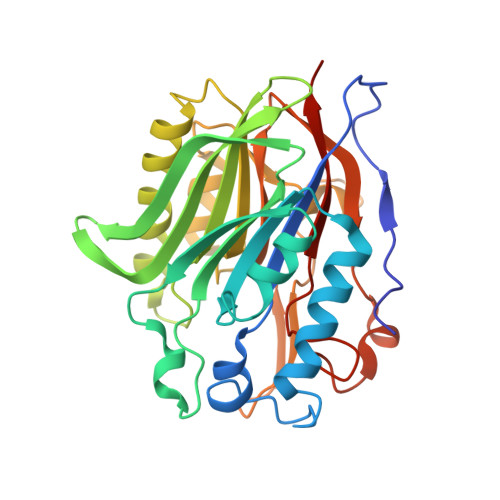
Crystal Structure of Human Nocturnin Catalytic Domain.

Estrella, M.A.Du, J.Korennykh, A.

(2018) Sci Rep 8: 16294-16294

  • DOI: https://doi.org/10.1038/s41598-018-34615-0
  • Primary Citation of Related Structures:  
    6MAL

  • PubMed Abstract: 

    Nocturnin (NOCT) helps the circadian clock to adjust metabolism according to day and night activity. NOCT is upregulated in early evening and it has been proposed that NOCT serves as a deadenylase for metabolic enzyme mRNAs. We present a 2.7-Å crystal structure of the catalytic domain of human NOCT. Our structure shows that NOCT has a close overall similarity to CCR4 deadenylase family members, PDE12 and CNOT6L, and to a DNA repair enzyme TDP2. All the key catalytic residues present in PDE12, CNOT6L and TDP2 are conserved in NOCT and have the same conformations. However, we observe substantial differences in the surface properties of NOCT, an unexpectedly narrow active site pocket, and conserved structural elements in the vicinity of the catalytic center, which are unique to NOCT and absent in the deadenylases PDE12/CNOT6L. Moreover, we show that in contrast to human PDE12 and CNOT6L, NOCT is completely inactive against poly-A RNA. Our work thus reveals the structure of an intriguing circadian protein and suggests that NOCT has considerable differences from the related deadenylases, which may point to a unique cellular function of this enzyme.


  • Organizational Affiliation

    Department of Molecular Biology, Princeton, NJ, 08544, USA.


Macromolecules
Find similar proteins by:  (by identity cutoff)  |  3D Structure
Entity ID: 1
MoleculeChains Sequence LengthOrganismDetailsImage
Nocturnin312Homo sapiensMutation(s): 0 
Gene Names: NOCTCCR4CCRN4LNOC
EC: 3.1.13.4
UniProt & NIH Common Fund Data Resources
Find proteins for Q9UK39 (Homo sapiens)
Explore Q9UK39 
Go to UniProtKB:  Q9UK39
PHAROS:  Q9UK39
GTEx:  ENSG00000151014 
Entity Groups  
Sequence Clusters30% Identity50% Identity70% Identity90% Identity95% Identity100% Identity
UniProt GroupQ9UK39
Sequence Annotations
Expand
  • Reference Sequence
Small Molecules
Ligands 1 Unique
IDChains Name / Formula / InChI Key2D Diagram3D Interactions
MG
Query on MG

Download Ideal Coordinates CCD File 
B [auth A]MAGNESIUM ION
Mg
JLVVSXFLKOJNIY-UHFFFAOYSA-N
Experimental Data & Validation

Experimental Data

  • Method: X-RAY DIFFRACTION
  • Resolution: 2.60 Å
  • R-Value Free: 0.307 
  • R-Value Work: 0.252 
  • R-Value Observed: 0.255 
  • Space Group: P 41 21 2
Unit Cell:
Length ( Å )Angle ( ˚ )
a = 61.4α = 90
b = 61.4β = 90
c = 155.3γ = 90
Software Package:
Software NamePurpose
PHENIXrefinement
XDSdata reduction
XSCALEdata scaling
PHASERphasing

Structure Validation

View Full Validation Report


Entry History & Funding Information

Deposition Data


Funding OrganizationLocationGrant Number
National Institutes of Health/National Institute of General Medical Sciences (NIH/NIGMS)United States1R01GM110161-01
Other privateUnited StatesAWD1004002
Other privateUnited States1013579

Revision History  (Full details and data files)

  • Version 1.0: 2018-09-05
    Type: Initial release
  • Version 1.1: 2018-11-21
    Changes: Data collection, Database references
  • Version 1.2: 2020-01-01
    Changes: Author supporting evidence
  • Version 1.3: 2023-10-11
    Changes: Data collection, Database references, Refinement description