6M5M

SPL-1 - GlcNAc complex


Experimental Data Snapshot

  • Method: X-RAY DIFFRACTION
  • Resolution: 1.70 Å
  • R-Value Free: 0.216 
  • R-Value Work: 0.203 
  • R-Value Observed: 0.204 

wwPDB Validation   3D Report Full Report


Ligand Structure Quality Assessment 


This is version 1.3 of the entry. See complete history


Literature

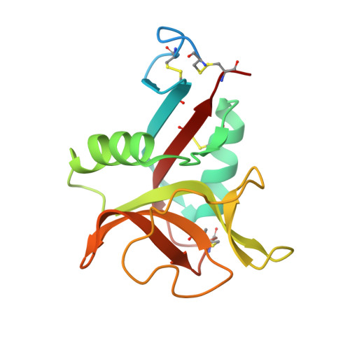
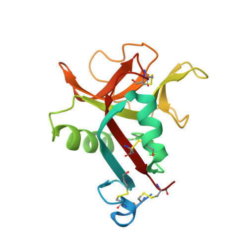
Novel carbohydrate-recognition mode of the invertebrate C-type lectin SPL-1 from Saxidomus purpuratus revealed by the GlcNAc-complex crystal in the presence of Ca2.

Unno, H.Higuchi, S.Goda, S.Hatakeyama, T.

(2020) Acta Crystallogr F Struct Biol Commun 76: 271-277

  • DOI: https://doi.org/10.1107/S2053230X20007256
  • Primary Citation of Related Structures:  
    6M5M

  • PubMed Abstract: 

    The C-type lectins SPL-1 and SPL-2 from the bivalve Saxidomus purpuratus are composed of A and B chains and of two B chains, respectively. They bind specific carbohydrates containing acetamido groups, such as N-acetylglucosamine (GlcNAc) and N-acetylgalactosamine (GalNAc), in a Ca 2+ -independent manner. Unlike ordinary C-type lectins, which require Ca 2+ ions for carbohydrate recognition, these lectins recognize specific carbohydrates mainly through interactions with the acetamido group without Ca 2+ ions, even though Ca 2+ enhances the binding affinity of these lectins, especially SPL-1. In the present study, the crystal structure of the SPL-1-GlcNAc complex in the presence of Ca 2+ revealed that the binding of SPL-1 to GlcNAc is stabilized by hydrogen bonds to the water molecule(s) coordinating Ca 2+ , whereas in ordinary C-type lectins Ca 2+ directly forms coordinate bonds to the hydroxy groups of carbohydrates. These differences may also allow SPL-1 and SPL-2 to recognize both GlcNAc and GalNAc, which have different orientations of the 4-hydroxy group.


  • Organizational Affiliation

    Biomolecular Chemistry Laboratory, Graduate School of Engineering, Nagasaki University, 1-14 Bunkyo-machi, Nagasaki 852-8521, Japan.


Macromolecules
Find similar proteins by:  (by identity cutoff)  |  3D Structure
Entity ID: 1
MoleculeChains Sequence LengthOrganismDetailsImage
N-acetylglucosamine-specific lectin158Saxidomus purpurataMutation(s): 0 
UniProt
Find proteins for A0A2Z6G7U6 (Saxidomus purpurata)
Explore A0A2Z6G7U6 
Go to UniProtKB:  A0A2Z6G7U6
Entity Groups  
Sequence Clusters30% Identity50% Identity70% Identity90% Identity95% Identity100% Identity
UniProt GroupA0A2Z6G7U6
Sequence Annotations
Expand
  • Reference Sequence
Find similar proteins by:  (by identity cutoff)  |  3D Structure
Entity ID: 2
MoleculeChains Sequence LengthOrganismDetailsImage
N-acetylglucosamine-specific lectin159Saxidomus purpurataMutation(s): 0 
UniProt
Find proteins for A0A2Z6G7U3 (Saxidomus purpurata)
Explore A0A2Z6G7U3 
Go to UniProtKB:  A0A2Z6G7U3
Entity Groups  
Sequence Clusters30% Identity50% Identity70% Identity90% Identity95% Identity100% Identity
UniProt GroupA0A2Z6G7U3
Sequence Annotations
Expand
  • Reference Sequence
Experimental Data & Validation

Experimental Data

  • Method: X-RAY DIFFRACTION
  • Resolution: 1.70 Å
  • R-Value Free: 0.216 
  • R-Value Work: 0.203 
  • R-Value Observed: 0.204 
  • Space Group: P 65
Unit Cell:
Length ( Å )Angle ( ˚ )
a = 44.46α = 90
b = 44.46β = 90
c = 303.613γ = 120
Software Package:
Software NamePurpose
Aimlessdata scaling
REFMACrefinement
PDB_EXTRACTdata extraction
XDSdata reduction
PHASERphasing

Structure Validation

View Full Validation Report


Ligand Structure Quality Assessment 


Entry History 

Deposition Data

Revision History  (Full details and data files)

  • Version 1.0: 2020-06-10
    Type: Initial release
  • Version 1.1: 2020-07-08
    Changes: Database references
  • Version 1.2: 2020-07-29
    Type: Remediation
    Reason: Carbohydrate remediation
    Changes: Data collection, Derived calculations, Structure summary
  • Version 1.3: 2023-11-29
    Changes: Data collection, Database references, Refinement description, Structure summary