6M3G

Crystal structure of human HPF1


Experimental Data Snapshot

  • Method: X-RAY DIFFRACTION
  • Resolution: 1.57 Å
  • R-Value Free: 0.205 
  • R-Value Work: 0.188 
  • R-Value Observed: 0.189 

wwPDB Validation   3D Report Full Report


This is version 1.1 of the entry. See complete history


Literature

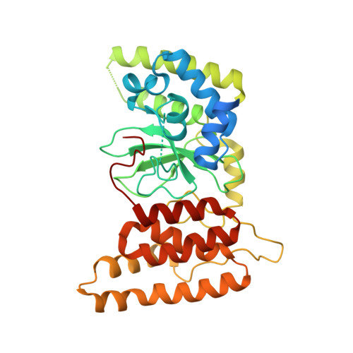
HPF1 remodels the active site of PARP1 to enable the serine ADP-ribosylation of histones.

Sun, F.H.Zhao, P.Zhang, N.Kong, L.L.Wong, C.C.L.Yun, C.H.

(2021) Nat Commun 12: 1028-1028

  • DOI: https://doi.org/10.1038/s41467-021-21302-4
  • Primary Citation of Related Structures:  
    6M3G, 6M3H, 6M3I

  • PubMed Abstract: 

    Upon binding to DNA breaks, poly(ADP-ribose) polymerase 1 (PARP1) ADP-ribosylates itself and other factors to initiate DNA repair. Serine is the major residue for ADP-ribosylation upon DNA damage, which strictly depends on HPF1. Here, we report the crystal structures of human HPF1/PARP1-CAT ΔHD complex at 1.98 Å resolution, and mouse and human HPF1 at 1.71 Å and 1.57 Å resolution, respectively. Our structures and mutagenesis data confirm that the structural insights obtained in a recent HPF1/PARP2 study by Suskiewicz et al. apply to PARP1. Moreover, we quantitatively characterize the key residues necessary for HPF1/PARP1 binding. Our data show that through salt-bridging to Glu284/Asp286, Arg239 positions Glu284 to catalyze serine ADP-ribosylation, maintains the local conformation of HPF1 to limit PARP1 automodification, and facilitates HPF1/PARP1 binding by neutralizing the negative charge of Glu284. These findings, along with the high-resolution structural data, may facilitate drug discovery targeting PARP1.


  • Organizational Affiliation

    Department of Biochemistry and Biophysics, Peking University Health Science Center, Beijing, China.


Macromolecules
Find similar proteins by:  (by identity cutoff)  |  3D Structure
Entity ID: 1
MoleculeChains Sequence LengthOrganismDetailsImage
Histone PARylation factor 1346Homo sapiensMutation(s): 0 
Gene Names: HPF1C4orf27
UniProt & NIH Common Fund Data Resources
Find proteins for Q9NWY4 (Homo sapiens)
Explore Q9NWY4 
Go to UniProtKB:  Q9NWY4
PHAROS:  Q9NWY4
GTEx:  ENSG00000056050 
Entity Groups  
Sequence Clusters30% Identity50% Identity70% Identity90% Identity95% Identity100% Identity
UniProt GroupQ9NWY4
Sequence Annotations
Expand
  • Reference Sequence
Experimental Data & Validation

Experimental Data

  • Method: X-RAY DIFFRACTION
  • Resolution: 1.57 Å
  • R-Value Free: 0.205 
  • R-Value Work: 0.188 
  • R-Value Observed: 0.189 
  • Space Group: H 3 2
Unit Cell:
Length ( Å )Angle ( ˚ )
a = 89.981α = 90
b = 89.981β = 90
c = 226.88γ = 120
Software Package:
Software NamePurpose
PHENIXrefinement
HKL-3000data reduction
HKL-3000data scaling
SHARPphasing

Structure Validation

View Full Validation Report


Entry History & Funding Information

Deposition Data


Funding OrganizationLocationGrant Number
National Natural Science Foundation of China (NSFC)China31270769

Revision History  (Full details and data files)

  • Version 1.0: 2021-03-03
    Type: Initial release
  • Version 1.1: 2024-03-27
    Changes: Data collection, Database references