6LL8

Type II inorganic pyrophosphatase (PPase) from the psychrophilic bacterium Shewanella sp. AS-11, Mg-PNP form


Experimental Data Snapshot

  • Method: X-RAY DIFFRACTION
  • Resolution: 1.30 Å
  • R-Value Free: 0.122 
  • R-Value Work: 0.097 

wwPDB Validation   3D Report Full Report


Ligand Structure Quality Assessment 


This is version 1.1 of the entry. See complete history


Literature

X-ray Crystallography and Electron Paramagnetic Resonance Spectroscopy Reveal Active Site Rearrangement of Cold-Adapted Inorganic Pyrophosphatase.

Horitani, M.Kusubayashi, K.Oshima, K.Yato, A.Sugimoto, H.Watanabe, K.

(2020) Sci Rep 10: 4368-4368

  • DOI: https://doi.org/10.1038/s41598-020-61217-6
  • Primary Citation of Related Structures:  
    6LL7, 6LL8

  • PubMed Abstract: 

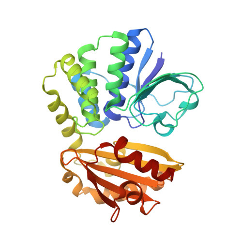
    Inorganic pyrophosphatase (PPase) catalyses the hydrolysis reaction of inorganic pyrophosphate to phosphates. Our previous studies showed that manganese (Mn) activated PPase from the psychrophilic bacterium Shewanella sp. AS-11 (Mn-Sh-PPase) has a characteristic temperature dependence of the activity with an optimum at 5 °C. Here we report the X-ray crystallography and electron paramagnetic resonance (EPR) spectroscopy structural analyses of Sh-PPase in the absence and presence of substrate analogues. We successfully determined the crystal structure of Mn-Sh-PPase without substrate and Mg-activated Sh-PPase (Mg-Sh-PPase) complexed with substrate analogue (imidodiphosphate; PNP). Crystallographic studies revealed a bridged water placed at a distance from the di-Mn centre in Mn-Sh-PPase without substrate. The water came closer to the metal centre when PNP bound. EPR analysis of Mn-Sh-PPase without substrate revealed considerably weak exchange coupling, whose magnitude was increased by binding of substrate analogues. The data indicate that the bridged molecule has weak bonds with the di-Mn centre, which suggests a 'loose' structure, whereas it comes closer to di-Mn centre by substrate binding, which suggests a 'well-tuned' structure for catalysis. Thus, we propose that Sh-PPase can rearrange the active site and that the 'loose' structure plays an important role in the cold adaptation mechanism.


  • Organizational Affiliation

    Department of Applied Biochemistry and Food Science, Saga University, 1 Honjo-machi, Saga, Saga, 840-8502, Japan. horitani@cc.saga-u.ac.jp.


Macromolecules
Find similar proteins by:  (by identity cutoff)  |  3D Structure
Entity ID: 1
MoleculeChains Sequence LengthOrganismDetailsImage
Inorganic pyrophosphatase
A, B
309Shewanella sp. AS-11Mutation(s): 0 
Gene Names: ppia
UniProt
Find proteins for L8AXY8 (Shewanella sp. AS-11)
Explore L8AXY8 
Go to UniProtKB:  L8AXY8
Entity Groups  
Sequence Clusters30% Identity50% Identity70% Identity90% Identity95% Identity100% Identity
UniProt GroupL8AXY8
Sequence Annotations
Expand
  • Reference Sequence
Small Molecules
Ligands 6 Unique
IDChains Name / Formula / InChI Key2D Diagram3D Interactions
EPE
Query on EPE

Download Ideal Coordinates CCD File 
J [auth A],
T [auth B]
4-(2-HYDROXYETHYL)-1-PIPERAZINE ETHANESULFONIC ACID
C8 H18 N2 O4 S
JKMHFZQWWAIEOD-UHFFFAOYSA-N
2PN (Subject of Investigation/LOI)
Query on 2PN

Download Ideal Coordinates CCD File 
I [auth A],
S [auth B]
IMIDODIPHOSPHORIC ACID
H5 N O6 P2
GNGSOPFGGKKDQP-UHFFFAOYSA-N
GOL
Query on GOL

Download Ideal Coordinates CCD File 
K [auth A],
L [auth A],
U [auth B],
V [auth B]
GLYCEROL
C3 H8 O3
PEDCQBHIVMGVHV-UHFFFAOYSA-N
CA
Query on CA

Download Ideal Coordinates CCD File 
H [auth A]CALCIUM ION
Ca
BHPQYMZQTOCNFJ-UHFFFAOYSA-N
MG
Query on MG

Download Ideal Coordinates CCD File 
C [auth A]
D [auth A]
E [auth A]
F [auth A]
G [auth A]
C [auth A],
D [auth A],
E [auth A],
F [auth A],
G [auth A],
N [auth B],
O [auth B],
P [auth B],
Q [auth B],
R [auth B]
MAGNESIUM ION
Mg
JLVVSXFLKOJNIY-UHFFFAOYSA-N
F
Query on F

Download Ideal Coordinates CCD File 
M [auth A],
W [auth B]
FLUORIDE ION
F
KRHYYFGTRYWZRS-UHFFFAOYSA-M
Experimental Data & Validation

Experimental Data

  • Method: X-RAY DIFFRACTION
  • Resolution: 1.30 Å
  • R-Value Free: 0.122 
  • R-Value Work: 0.097 
  • Space Group: P 1 21 1
Unit Cell:
Length ( Å )Angle ( ˚ )
a = 53.49α = 90
b = 78.98β = 98.593
c = 75.3γ = 90
Software Package:
Software NamePurpose
XDSdata scaling
REFMACrefinement
PDB_EXTRACTdata extraction
PHASERphasing
Cootmodel building
XDSdata reduction

Structure Validation

View Full Validation Report


Ligand Structure Quality Assessment 


Entry History & Funding Information

Deposition Data


Funding OrganizationLocationGrant Number
Japan Society for the Promotion of Science (JSPS)Japan17H06955
Japan Society for the Promotion of Science (JSPS)Japan18H02412

Revision History  (Full details and data files)

  • Version 1.0: 2020-03-25
    Type: Initial release
  • Version 1.1: 2023-11-22
    Changes: Data collection, Database references, Derived calculations, Refinement description