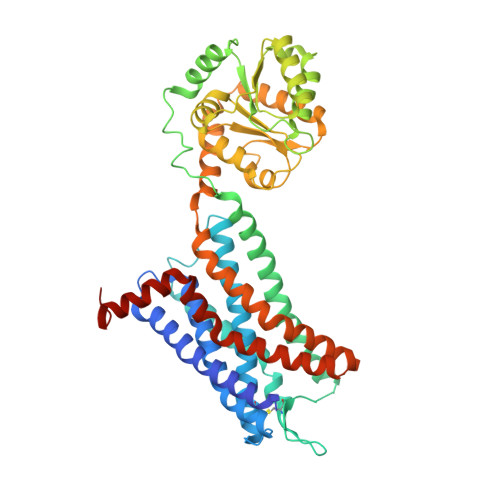
6LFL

Crystal structure of a class A GPCR


Experimental Data Snapshot

  • Method: X-RAY DIFFRACTION
  • Resolution: 3.20 Å
  • R-Value Free: 0.264 
  • R-Value Work: 0.231 
  • R-Value Observed: 0.233 

wwPDB Validation   3D Report Full Report


Ligand Structure Quality Assessment 


This is version 1.2 of the entry. See complete history


Literature

Structural basis of CXC chemokine receptor 2 activation and signalling.

Liu, K.Wu, L.Yuan, S.Wu, M.Xu, Y.Sun, Q.Li, S.Zhao, S.Hua, T.Liu, Z.J.

(2020) Nature 585: 135-140

  • DOI: https://doi.org/10.1038/s41586-020-2492-5
  • Primary Citation of Related Structures:  
    6LFL, 6LFM, 6LFO

  • PubMed Abstract: 

    Chemokines and their receptors mediate cell migration, which influences multiple fundamental biological processes and disease conditions such as inflammation and cancer 1 . Although ample effort has been invested into the structural investigation of the chemokine receptors and receptor-chemokine recognition 2-4 , less is known about endogenous chemokine-induced receptor activation and G-protein coupling. Here we present the cryo-electron microscopy structures of interleukin-8 (IL-8, also known as CXCL8)-activated human CXC chemokine receptor 2 (CXCR2) in complex with G i  protein, along with a crystal structure of CXCR2 bound to a designed allosteric antagonist. Our results reveal a unique shallow mode of binding between CXCL8 and CXCR2, and also show the interactions between CXCR2 and G i  protein. Further structural analysis of the inactive and active states of CXCR2 reveals a distinct activation process and the competitive small-molecule antagonism of chemokine receptors. In addition, our results provide insights into how a G-protein-coupled receptor is activated by an endogenous protein molecule, which will assist in the rational development of therapeutics that target the chemokine system for better pharmacological profiles.


  • Organizational Affiliation

    iHuman Institute, ShanghaiTech University, Shanghai, China.


Macromolecules
Find similar proteins by:  (by identity cutoff)  |  3D Structure
Entity ID: 1
MoleculeChains Sequence LengthOrganismDetailsImage
C-X-C chemokine receptor type 2,GlgA glycogen synthase,C-X-C chemokine receptor type 2502Homo sapiensPyrococcus abyssi GE5Mutation(s): 3 
Gene Names: CXCR2IL8RBPAB2292
Membrane Entity: Yes 
UniProt & NIH Common Fund Data Resources
Find proteins for Q9V2J8 (Pyrococcus abyssi (strain GE5 / Orsay))
Explore Q9V2J8 
Go to UniProtKB:  Q9V2J8
Find proteins for P25025 (Homo sapiens)
Explore P25025 
Go to UniProtKB:  P25025
PHAROS:  P25025
GTEx:  ENSG00000180871 
Entity Groups  
Sequence Clusters30% Identity50% Identity70% Identity90% Identity95% Identity100% Identity
UniProt GroupsQ9V2J8P25025
Sequence Annotations
Expand
  • Reference Sequence
Small Molecules
Ligands 1 Unique
IDChains Name / Formula / InChI Key2D Diagram3D Interactions
EBX (Subject of Investigation/LOI)
Query on EBX

Download Ideal Coordinates CCD File 
B [auth A]4-[[3,4-bis(oxidanylidene)-2-[[(1~{R})-1-(4-propan-2-ylfuran-2-yl)propyl]amino]cyclobuten-1-yl]amino]-~{N},~{N}-dimethyl-3-oxidanyl-pyridine-2-carboxamide
C22 H26 N4 O5
JSZQMZJVSCVDMA-CYBMUJFWSA-N
Experimental Data & Validation

Experimental Data

  • Method: X-RAY DIFFRACTION
  • Resolution: 3.20 Å
  • R-Value Free: 0.264 
  • R-Value Work: 0.231 
  • R-Value Observed: 0.233 
  • Space Group: P 1 21 1
Unit Cell:
Length ( Å )Angle ( ˚ )
a = 41.3α = 90
b = 108.46β = 91.16
c = 82.6γ = 90
Software Package:
Software NamePurpose
BUSTERrefinement
XDSdata reduction
XSCALEdata scaling
PHASERphasing
PDB_EXTRACTdata extraction

Structure Validation

View Full Validation Report


Ligand Structure Quality Assessment 


Entry History 

Deposition Data

Revision History  (Full details and data files)

  • Version 1.0: 2020-09-02
    Type: Initial release
  • Version 1.1: 2020-09-16
    Changes: Database references
  • Version 1.2: 2023-11-22
    Changes: Data collection, Database references, Refinement description