6LB7

Crystal structure of the Ca2+-free and Ca2+-bound MICU1-MICU2 complex


Experimental Data Snapshot

  • Method: X-RAY DIFFRACTION
  • Resolution: 2.10 Å
  • R-Value Free: 0.227 
  • R-Value Work: 0.194 
  • R-Value Observed: 0.195 

wwPDB Validation   3D Report Full Report


This is version 1.3 of the entry. See complete history


Literature

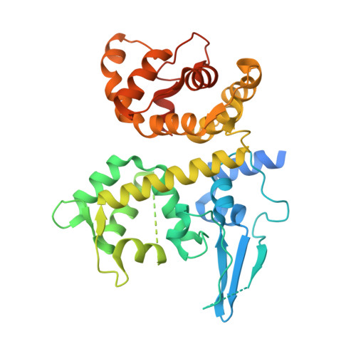
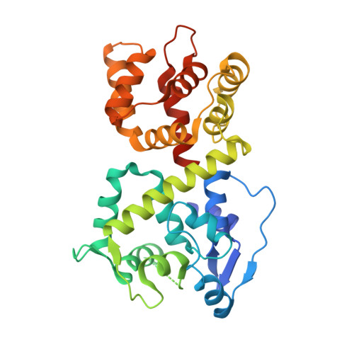
The structure of the MICU1-MICU2 complex unveils the regulation of the mitochondrial calcium uniporter.

Wu, W.Shen, Q.Zhang, R.Qiu, Z.Wang, Y.Zheng, J.Jia, Z.

(2020) EMBO J 39: e104285-e104285

  • DOI: https://doi.org/10.15252/embj.2019104285
  • Primary Citation of Related Structures:  
    6LB7, 6LB8

  • PubMed Abstract: 

    The MICU1-MICU2 heterodimer regulates the mitochondrial calcium uniporter (MCU) and mitochondrial calcium uptake. Herein, we present two crystal structures of the MICU1-MICU2 heterodimer, in which Ca 2+ -free and Ca 2+ -bound EF-hands are observed in both proteins, revealing both electrostatic and hydrophobic interfaces. Furthermore, we show that MICU1 interacts with EMRE, another regulator of MCU, through a Ca 2+ -dependent alkaline groove. Ca 2+ binding strengthens the MICU1-EMRE interaction, which in turn facilitates Ca 2+ uptake. Conversely, the MICU1-MCU interaction is favored in the absence of Ca 2+ , thus inhibiting the channel activity. This Ca 2+ -dependent switch illuminates how calcium signals are transmitted from regulatory subunits to the calcium channel and the transition between gatekeeping and activation channel functions. Furthermore, competition with an EMRE peptide alters the uniporter threshold in resting conditions and elevates Ca 2+ accumulation in stimulated mitochondria, confirming the gatekeeper role of the MICU1-MICU2 heterodimer. Taken together, these structural and functional data provide new insights into the regulation of mitochondrial calcium uptake.


  • Organizational Affiliation

    College of Chemistry, Beijing Normal University, Beijing, China.


Macromolecules
Find similar proteins by:  (by identity cutoff)  |  3D Structure
Entity ID: 1
MoleculeChains Sequence LengthOrganismDetailsImage
Calcium uptake protein 1, mitochondrial
A, C
377Homo sapiensMutation(s): 0 
Gene Names: MICU1CALCCBARA1
UniProt & NIH Common Fund Data Resources
Find proteins for Q9BPX6 (Homo sapiens)
Explore Q9BPX6 
Go to UniProtKB:  Q9BPX6
PHAROS:  Q9BPX6
GTEx:  ENSG00000107745 
Entity Groups  
Sequence Clusters30% Identity50% Identity70% Identity90% Identity95% Identity100% Identity
UniProt GroupQ9BPX6
Sequence Annotations
Expand
  • Reference Sequence
Find similar proteins by:  (by identity cutoff)  |  3D Structure
Entity ID: 2
MoleculeChains Sequence LengthOrganismDetailsImage
Calcium uptake protein 2, mitochondrial
B, D
330Homo sapiensMutation(s): 0 
Gene Names: MICU2EFHA1
UniProt & NIH Common Fund Data Resources
Find proteins for Q8IYU8 (Homo sapiens)
Explore Q8IYU8 
Go to UniProtKB:  Q8IYU8
PHAROS:  Q8IYU8
GTEx:  ENSG00000165487 
Entity Groups  
Sequence Clusters30% Identity50% Identity70% Identity90% Identity95% Identity100% Identity
UniProt GroupQ8IYU8
Sequence Annotations
Expand
  • Reference Sequence
Experimental Data & Validation

Experimental Data

  • Method: X-RAY DIFFRACTION
  • Resolution: 2.10 Å
  • R-Value Free: 0.227 
  • R-Value Work: 0.194 
  • R-Value Observed: 0.195 
  • Space Group: P 21 21 21
Unit Cell:
Length ( Å )Angle ( ˚ )
a = 85.481α = 90
b = 107.721β = 90
c = 170.381γ = 90
Software Package:
Software NamePurpose
PHENIXrefinement
HKL-2000data scaling
PDB_EXTRACTdata extraction
HKL-2000data reduction
PHENIXphasing

Structure Validation

View Full Validation Report


Entry History & Funding Information

Deposition Data


Funding OrganizationLocationGrant Number
National Natural Science Foundation of ChinaChina21773014

Revision History  (Full details and data files)

  • Version 1.0: 2020-07-15
    Type: Initial release
  • Version 1.1: 2021-01-27
    Changes: Database references, Derived calculations
  • Version 1.2: 2021-03-17
    Changes: Database references
  • Version 1.3: 2023-11-22
    Changes: Data collection, Database references, Refinement description