6L8S

High resolution crystal structure of crustacean hemocyanin.


Experimental Data Snapshot

  • Method: X-RAY DIFFRACTION
  • Resolution: 1.58 Å
  • R-Value Free: 0.186 
  • R-Value Work: 0.149 
  • R-Value Observed: 0.151 

wwPDB Validation   3D Report Full Report


This is version 1.3 of the entry. See complete history


Literature

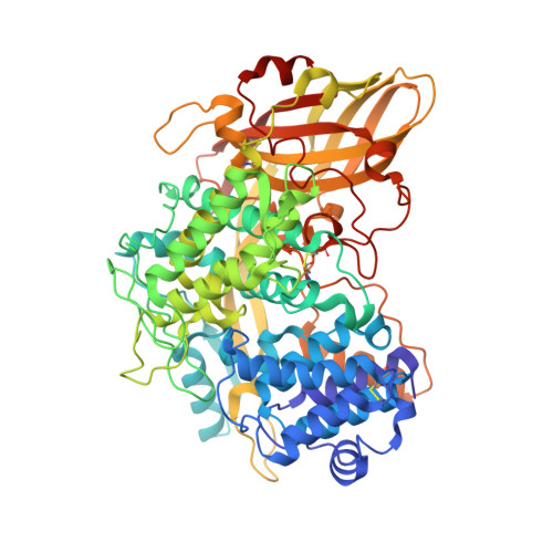
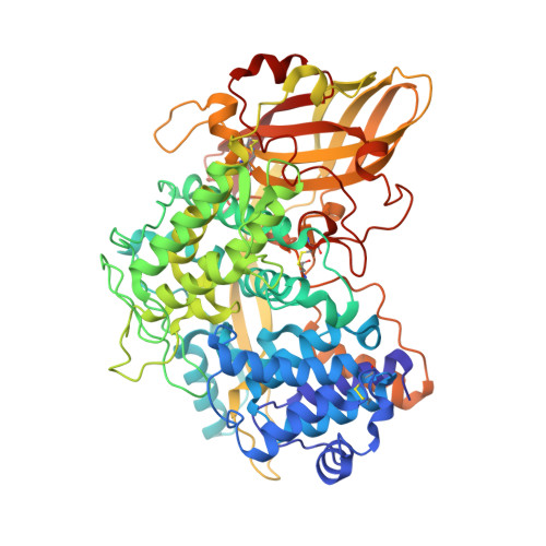
The high-resolution crystal structure of lobster hemocyanin shows its enzymatic capability as a phenoloxidase.

Masuda, T.Baba, S.Matsuo, K.Ito, S.Mikami, B.

(2020) Arch Biochem Biophys 688: 108370-108370

  • DOI: https://doi.org/10.1016/j.abb.2020.108370
  • Primary Citation of Related Structures:  
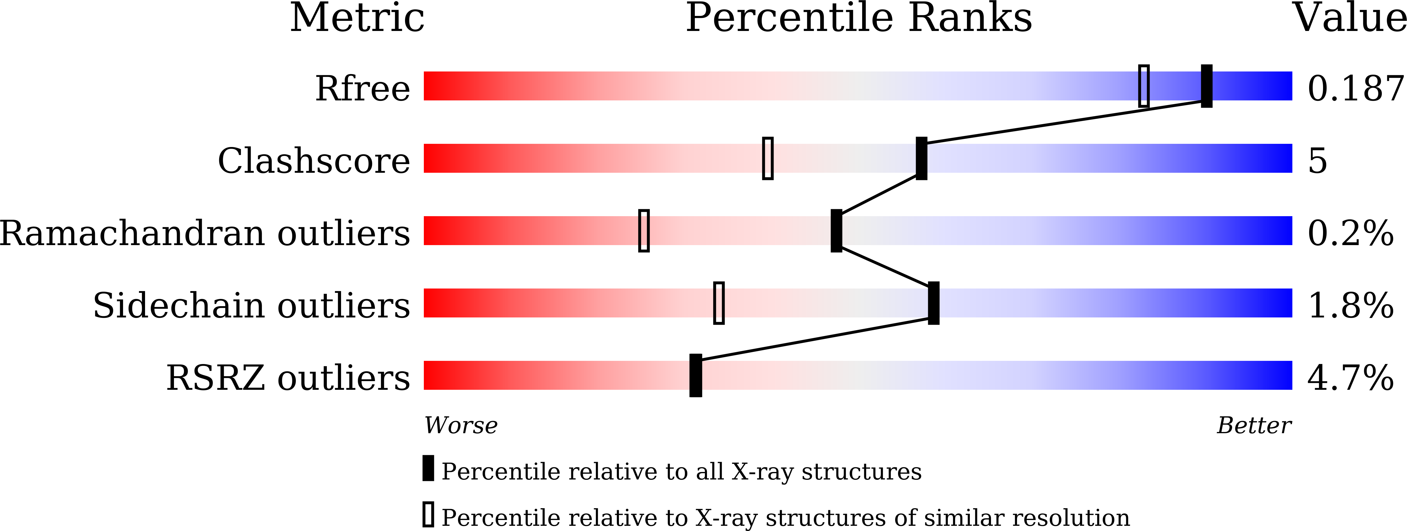
    6L8S

  • PubMed Abstract: 

    Hemocyanin (Hc) and phenoloxidase (PO) are members of the type 3 copper protein family. Although arthropod Hc and PO exhibit similar three-dimensional structures of the copper-containing active site, Hc functions as an oxygen transport protein, showing minimal or no phenoloxidase activity. Here, we present the crystal structure of the oxy form of Hc from Panulirus japonicus (PjHc) at 1.58 Å resolution. The structure of the di-copper active site of PjHc was found to be almost identical to that of PO. Although conserved amino acids and the water molecule crucial for the enzymatic activity were observed in PjHc at almost the same positions as those in PO, PjHc showed no enzymatic activity under our experimental conditions. One striking difference between PjHc and arthropod PO was the presence of a "blocker residue" near the binuclear copper site of PjHc. This blocker residue comprised a phenylalanine residue tightly stacked with an imidazole ring of a CuA coordinated histidine and hindered substrates from accessing the active site. Our results suggest that the blocker residue is also a determining factor of the catalytic activity of type 3 copper proteins.


  • Organizational Affiliation

    Laboratory of Marine Biology, Division of Applied Biological Science, Faculty of Agriculture, Setsunan University, 45-1 Nagaotoge-cho, Hirakata, Osaka, 573-0101, Japan. Electronic address: taro.masuda@setsunan.ac.jp.


Macromolecules
Find similar proteins by:  (by identity cutoff)  |  3D Structure
Entity ID: 1
MoleculeChains Sequence LengthOrganismDetailsImage
Hemocyanin650Panulirus japonicusMutation(s): 0 
Entity Groups  
Sequence Clusters30% Identity50% Identity70% Identity90% Identity95% Identity100% Identity
Sequence Annotations
Expand
  • Reference Sequence
Find similar proteins by:  (by identity cutoff)  |  3D Structure
Entity ID: 2
MoleculeChains Sequence LengthOrganismDetailsImage
Hemocyanin650Panulirus japonicusMutation(s): 0 
Entity Groups  
Sequence Clusters30% Identity50% Identity70% Identity90% Identity95% Identity100% Identity
Sequence Annotations
Expand
  • Reference Sequence
Find similar proteins by:  (by identity cutoff)  |  3D Structure
Entity ID: 3
MoleculeChains Sequence LengthOrganismDetailsImage
Hemocyanin650Panulirus japonicusMutation(s): 0 
Entity Groups  
Sequence Clusters30% Identity50% Identity70% Identity90% Identity95% Identity100% Identity
Sequence Annotations
Expand
  • Reference Sequence
Small Molecules
Ligands 7 Unique
IDChains Name / Formula / InChI Key2D Diagram3D Interactions
NAG
Query on NAG

Download Ideal Coordinates CCD File 
F [auth A],
R [auth B]
2-acetamido-2-deoxy-beta-D-glucopyranose
C8 H15 N O6
OVRNDRQMDRJTHS-FMDGEEDCSA-N
SO4
Query on SO4

Download Ideal Coordinates CCD File 
I [auth A],
IA [auth C],
U [auth B]
SULFATE ION
O4 S
QAOWNCQODCNURD-UHFFFAOYSA-L
CU (Subject of Investigation/LOI)
Query on CU

Download Ideal Coordinates CCD File 
D [auth A]
E [auth A]
EA [auth C]
FA [auth C]
P [auth B]
D [auth A],
E [auth A],
EA [auth C],
FA [auth C],
P [auth B],
Q [auth B]
COPPER (II) ION
Cu
JPVYNHNXODAKFH-UHFFFAOYSA-N
EDO
Query on EDO

Download Ideal Coordinates CCD File 
AA [auth B]
BA [auth B]
CA [auth B]
K [auth A]
L [auth A]
AA [auth B],
BA [auth B],
CA [auth B],
K [auth A],
L [auth A],
LA [auth C],
M [auth A],
MA [auth C],
N [auth A],
NA [auth C],
O [auth A],
OA [auth C],
PA [auth C],
QA [auth C],
X [auth B],
Y [auth B],
Z [auth B]
1,2-ETHANEDIOL
C2 H6 O2
LYCAIKOWRPUZTN-UHFFFAOYSA-N
CL
Query on CL

Download Ideal Coordinates CCD File 
DA [auth B]CHLORIDE ION
Cl
VEXZGXHMUGYJMC-UHFFFAOYSA-M
MG
Query on MG

Download Ideal Coordinates CCD File 
J [auth A],
JA [auth C],
KA [auth C],
V [auth B],
W [auth B]
MAGNESIUM ION
Mg
JLVVSXFLKOJNIY-UHFFFAOYSA-N
O
Query on O

Download Ideal Coordinates CCD File 
G [auth A]
GA [auth C]
H [auth A]
HA [auth C]
S [auth B]
G [auth A],
GA [auth C],
H [auth A],
HA [auth C],
S [auth B],
T [auth B]
OXYGEN ATOM
O
XLYOFNOQVPJJNP-UHFFFAOYSA-N
Experimental Data & Validation

Experimental Data

  • Method: X-RAY DIFFRACTION
  • Resolution: 1.58 Å
  • R-Value Free: 0.186 
  • R-Value Work: 0.149 
  • R-Value Observed: 0.151 
  • Space Group: C 2 2 21
Unit Cell:
Length ( Å )Angle ( ˚ )
a = 119.454α = 90
b = 207.596β = 90
c = 187.364γ = 90
Software Package:
Software NamePurpose
XDSdata reduction
XSCALEdata scaling
PHENIXrefinement
PDB_EXTRACTdata extraction
MOLREPphasing

Structure Validation

View Full Validation Report


Entry History & Funding Information

Deposition Data


Funding OrganizationLocationGrant Number
Japan Society for the Promotion of ScienceJapan19K06221

Revision History  (Full details and data files)

  • Version 1.0: 2020-05-27
    Type: Initial release
  • Version 1.1: 2020-06-17
    Changes: Database references
  • Version 1.2: 2020-07-29
    Type: Remediation
    Reason: Carbohydrate remediation
    Changes: Data collection, Derived calculations, Structure summary
  • Version 1.3: 2023-11-22
    Changes: Data collection, Database references, Refinement description, Structure summary