


Crystal structure of Hexokinase from Eimeria tenella
Yuan, H., Sun, M.F., Wang, Y.H., Liao, S.Q.To be published.
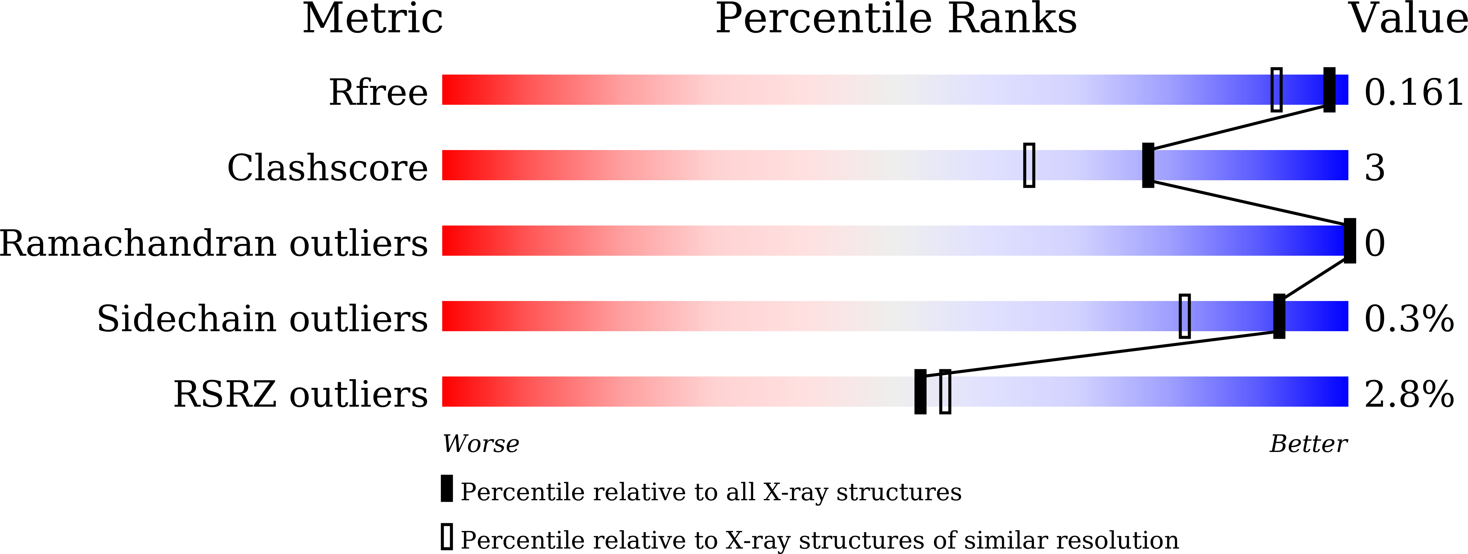
Experimental Data Snapshot
Entity ID: 1 | |||||
|---|---|---|---|---|---|
| Molecule | Chains | Sequence Length | Organism | Details | Image |
| Phosphotransferase | 467 | Eimeria tenella | Mutation(s): 0 EC: 2.7.1 | 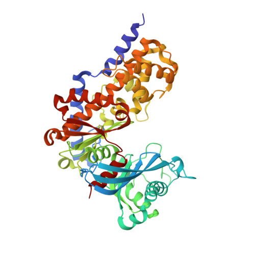 | |
UniProt | |||||
Find proteins for U6KUE1 (Eimeria tenella) Explore U6KUE1 Go to UniProtKB: U6KUE1 | |||||
Entity Groups | |||||
| Sequence Clusters | 30% Identity50% Identity70% Identity90% Identity95% Identity100% Identity | ||||
| UniProt Group | U6KUE1 | ||||
Sequence AnnotationsExpand | |||||
| |||||
| Ligands 1 Unique | |||||
|---|---|---|---|---|---|
| ID | Chains | Name / Formula / InChI Key | 2D Diagram | 3D Interactions | |
| GLA (Subject of Investigation/LOI) Query on GLA | B [auth A] | alpha-D-galactopyranose C6 H12 O6 WQZGKKKJIJFFOK-PHYPRBDBSA-N |  | ||
| Length ( Å ) | Angle ( ˚ ) |
|---|---|
| a = 68 | α = 90 |
| b = 77.05 | β = 90 |
| c = 105.15 | γ = 90 |
| Software Name | Purpose |
|---|---|
| PHENIX | refinement |
| HKL-3000 | data reduction |
| HKL-3000 | data scaling |
| MOLREP | phasing |
RCSB PDB (citation) is hosted by
RCSB PDB is a member of the

