6KLR

Crystal structure of human WIPI3 in complex with the WIR-peptide from ATG2A


Experimental Data Snapshot

  • Method: X-RAY DIFFRACTION
  • Resolution: 2.21 Å
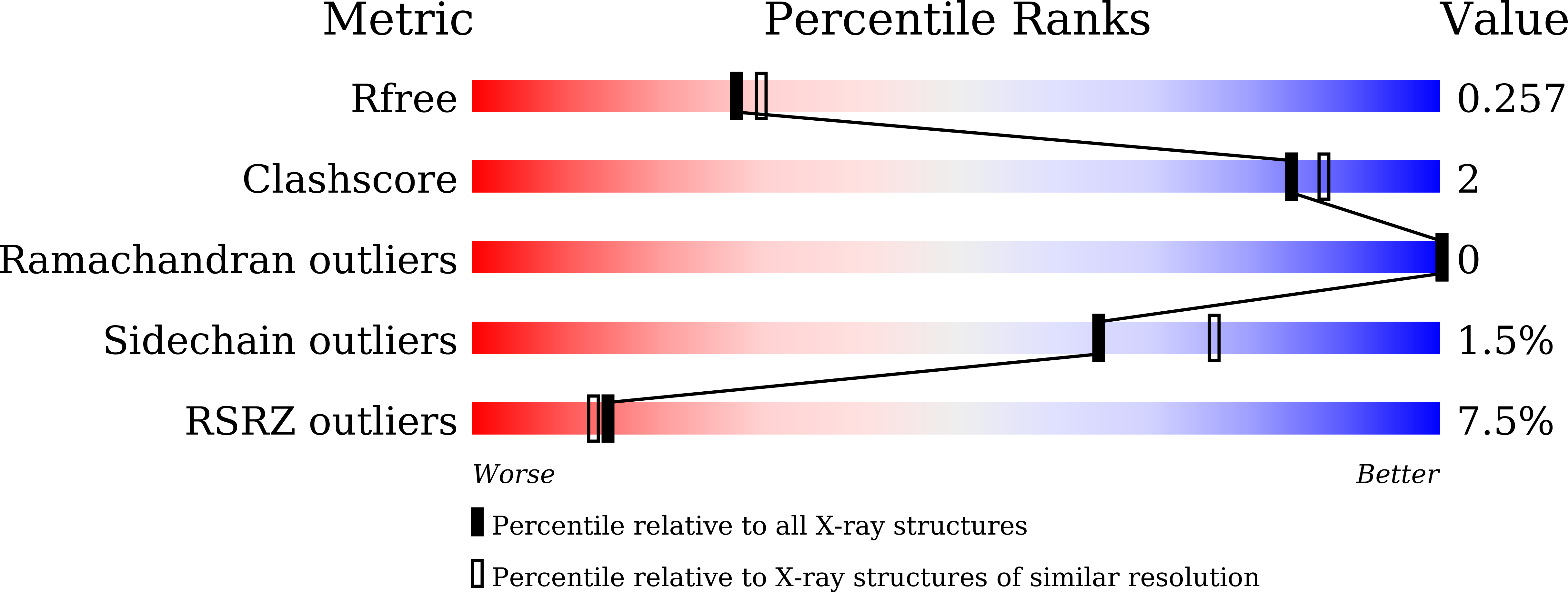
  • R-Value Free: 0.255 
  • R-Value Work: 0.213 
  • R-Value Observed: 0.215 

wwPDB Validation   3D Report Full Report


This is version 1.2 of the entry. See complete history


Literature

Multi-site-mediated entwining of the linear WIR-motif around WIPI beta-propellers for autophagy.

Ren, J.Liang, R.Wang, W.Zhang, D.Yu, L.Feng, W.

(2020) Nat Commun 11: 2702-2702

  • DOI: https://doi.org/10.1038/s41467-020-16523-y
  • Primary Citation of Related Structures:  
    6KLR

  • PubMed Abstract: 

    WIPI proteins (WIPI1-4) are mammalian PROPPIN family phosphoinositide effectors essential for autophagosome biogenesis. In addition to phosphoinositides, WIPI proteins can recognize a linear WIPI-interacting-region (WIR)-motif, but the underlying mechanism is poorly understood. Here, we determine the structure of WIPI3 in complex with the WIR-peptide from ATG2A. Unexpectedly, the WIR-peptide entwines around the WIPI3 seven-bladed β-propeller and binds to three sites in blades 1-3. The N-terminal part of the WIR-peptide forms a short strand that augments the periphery of blade 2, the middle segment anchors into an inter-blade hydrophobic pocket between blades 2-3, and the C-terminal aromatic tail wedges into another tailored pocket between blades 1-2. Mutations in three peptide-binding sites disrupt the interactions between WIPI3/4 and ATG2A and impair the ATG2A-mediated autophagic process. Thus, WIPI proteins recognize the WIR-motif by multi-sites in multi-blades and this multi-site-mediated peptide-recognition mechanism could be applicable to other PROPPIN proteins.


  • Organizational Affiliation

    National Laboratory of Biomacromolecules, CAS Center for Excellence in Biomacromolecules, Institute of Biophysics, Chinese Academy of Sciences, 15 Datun Road, 100101, Beijing, China.


Macromolecules
Find similar proteins by:  (by identity cutoff)  |  3D Structure
Entity ID: 1
MoleculeChains Sequence LengthOrganismDetailsImage
chimera ATG2A and WIPI3
A, B
350Homo sapiensMutation(s): 0 
Gene Names: ATG2AKIAA0404WDR45BWDR45LWIPI3
UniProt & NIH Common Fund Data Resources
Find proteins for Q2TAZ0 (Homo sapiens)
Explore Q2TAZ0 
Go to UniProtKB:  Q2TAZ0
PHAROS:  Q2TAZ0
GTEx:  ENSG00000110046 
Find proteins for Q5MNZ6 (Homo sapiens)
Explore Q5MNZ6 
Go to UniProtKB:  Q5MNZ6
PHAROS:  Q5MNZ6
GTEx:  ENSG00000141580 
Entity Groups  
Sequence Clusters30% Identity50% Identity70% Identity90% Identity95% Identity100% Identity
UniProt GroupsQ2TAZ0Q5MNZ6
Sequence Annotations
Expand
  • Reference Sequence
Experimental Data & Validation

Experimental Data

  • Method: X-RAY DIFFRACTION
  • Resolution: 2.21 Å
  • R-Value Free: 0.255 
  • R-Value Work: 0.213 
  • R-Value Observed: 0.215 
  • Space Group: P 21 21 2
Unit Cell:
Length ( Å )Angle ( ˚ )
a = 75.124α = 90
b = 121.063β = 90
c = 37.768γ = 90
Software Package:
Software NamePurpose
PHENIXrefinement
PDB_EXTRACTdata extraction
HKL-2000data reduction
HKL-2000data scaling
PHASERphasing

Structure Validation

View Full Validation Report


Entry History 

Deposition Data

Revision History  (Full details and data files)

  • Version 1.0: 2020-04-29
    Type: Initial release
  • Version 1.1: 2020-06-17
    Changes: Database references
  • Version 1.2: 2023-11-22
    Changes: Data collection, Database references, Refinement description