Crystallographic and calorimetric analysis on Pleurotus ostreatus lectin and its sugar complexes - promiscuous binding driven by geometry.
Vajravijayan, S., Pletnev, S., Luo, Z., Pletnev, V.Z., Nandhagopal, N., Gunasekaran, K.(2020) Int J Biol Macromol 152: 862-872
- PubMed: 32112837
- DOI: https://doi.org/10.1016/j.ijbiomac.2020.02.294
- Primary Citation of Related Structures:
6KBJ, 6KBQ, 6KC2, 6LI7, 6LIK - PubMed Abstract:
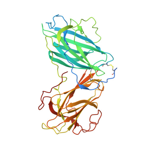
Carbohydrate recognition is established as a property of lectins and implicated in many functions including immunity and defense against pathogens. Many lectins are characterized and proposed for various applications owing to the above said recognition. The crystal structure of a lectin from Pleurotus ostreatus has been determined and shown to be calcium dependent. The overall structure is a tandem repeat of two β-jelly roll domains, a new fold for lectins. The calcium dependence of sugar binding is analyzed in-detail through isothermal titration calorimetry. The serendipitous observation of malonate and glycerol, the intentional N-Acetyl-D-galactosamine, D-Galactose and L-Rhamnose binding to Pleurotus ostreatus lectin by Ca 2+ coordination revealed that the binding site is promiscuous. Among these sugars, Rhamnose binding found to be thermodynamically most favourable. In all these structures, a vicinal diol motif, one at axial and the other at equatorial positions could be established as a specific requirement for binding. Interestingly, when compared with other calcium mediated lectin structures; this geometric requirement is found conserved. This observation could lead to the conclusion that lectins are not 'molecule specific' but 'geometry specific' so that any molecule not necessarily a sugar may be recognized by this lectin if the geometry exists.
Organizational Affiliation:
Centre of Advanced Study in Crystallography and Biophysics, University of Madras, Guindy Campus, Chennai 600 025, India.