6JZK

Structure of FimA type-1 (FimA1) prepilin of the type V major fimbrium


Experimental Data Snapshot

  • Method: X-RAY DIFFRACTION
  • Resolution: 2.10 Å
  • R-Value Free: 0.240 
  • R-Value Work: 0.189 
  • R-Value Observed: 0.192 

wwPDB Validation   3D Report Full Report


Ligand Structure Quality Assessment 


This is version 1.3 of the entry. See complete history


Literature

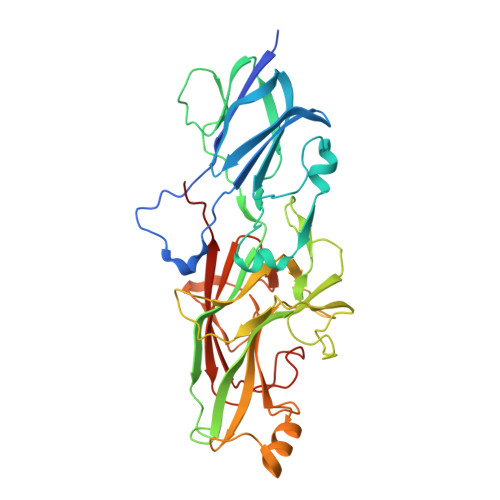
Structure of polymerized type V pilin reveals assembly mechanism involving protease-mediated strand exchange.

Shibata, S.Shoji, M.Okada, K.Matsunami, H.Matthews, M.M.Imada, K.Nakayama, K.Wolf, M.

(2020) Nat Microbiol 5: 830-837

  • DOI: https://doi.org/10.1038/s41564-020-0705-1
  • Primary Citation of Related Structures:  
    6JZJ, 6JZK, 6KMF

  • PubMed Abstract: 

    Bacterial adhesion is a general strategy for host-microbe and microbe-microbe interactions. Adhesive pili are essential for colonization, biofilm formation, virulence and pathogenesis of many environmental and pathogenic bacteria 1,2 . Members of the class Bacteroidia have unique type V pili, assembled by protease-mediated polymerization 3 . Porphyromonas gingivalis is the main contributor to periodontal disease and its type V pili are a key factor for its virulence 4 . However, the structure of the polymerized pilus and its assembly mechanism are unknown. Here we show structures of polymerized and monomeric states of FimA stalk pilin from P. gingivalis, determined by cryo-electron microscopy and crystallography. The atomic model of assembled FimA shows that the C-terminal strand of a donor subunit is inserted into a groove in the β-sheet of an acceptor subunit after N-terminal cleavage by the protease RgpB. The C terminus of the donor strand is essential for polymerization. We propose that type V pili assemble via a sequential polar assembly mechanism at the cell surface, involving protease-mediated strand exchange, employed by various Gram-negative species belonging to the class Bacteroidia. Our results reveal functional surfaces related to pathogenic properties of polymerized FimA. These insights may facilitate development of antibacterial drugs.


  • Organizational Affiliation

    Molecular Cryo-Electron Microscopy Unit, Okinawa Institute of Science and Technology Graduate University, Onna-son, Japan.


Macromolecules
Find similar proteins by:  (by identity cutoff)  |  3D Structure
Entity ID: 1
MoleculeChains Sequence LengthOrganismDetailsImage
Major fimbrium subunit FimA type-1
A, B
367Porphyromonas gingivalis ATCC 33277Mutation(s): 0 
Gene Names: fimAPGN_0180
UniProt
Find proteins for B2RH54 (Porphyromonas gingivalis (strain ATCC 33277 / DSM 20709 / CIP 103683 / JCM 12257 / NCTC 11834 / 2561))
Explore B2RH54 
Go to UniProtKB:  B2RH54
Entity Groups  
Sequence Clusters30% Identity50% Identity70% Identity90% Identity95% Identity100% Identity
UniProt GroupB2RH54
Sequence Annotations
Expand
  • Reference Sequence
Experimental Data & Validation

Experimental Data

  • Method: X-RAY DIFFRACTION
  • Resolution: 2.10 Å
  • R-Value Free: 0.240 
  • R-Value Work: 0.189 
  • R-Value Observed: 0.192 
  • Space Group: P 21 21 21
Unit Cell:
Length ( Å )Angle ( ˚ )
a = 35.84α = 90
b = 85.452β = 90
c = 242.561γ = 90
Software Package:
Software NamePurpose
PHENIXrefinement
MOSFLMdata reduction
Aimlessdata scaling
PHENIXphasing

Structure Validation

View Full Validation Report


Ligand Structure Quality Assessment 


Entry History & Funding Information

Deposition Data


Funding OrganizationLocationGrant Number
Japan Society for the Promotion of ScienceJapan16H05504

Revision History  (Full details and data files)

  • Version 1.0: 2020-04-15
    Type: Initial release
  • Version 1.1: 2020-04-29
    Changes: Database references
  • Version 1.2: 2020-06-17
    Changes: Database references
  • Version 1.3: 2023-11-22
    Changes: Data collection, Database references, Refinement description