6JFV

The crystal structure of 2B-2B complex from keratins 5 and 14 (C367A mutant of K14)


Experimental Data Snapshot

  • Method: X-RAY DIFFRACTION
  • Resolution: 2.60 Å
  • R-Value Free: 0.246 
  • R-Value Work: 0.201 
  • R-Value Observed: 0.203 

wwPDB Validation   3D Report Full Report


This is version 1.2 of the entry. See complete history


Literature

Structure-Function Analyses of a Keratin Heterotypic Complex Identify Specific Keratin Regions Involved in Intermediate Filament Assembly.

Lee, C.H.Kim, M.S.Li, S.Leahy, D.J.Coulombe, P.A.

(2020) Structure 28: 355-362.e4

  • DOI: https://doi.org/10.1016/j.str.2020.01.002
  • Primary Citation of Related Structures:  
    6JFV

  • PubMed Abstract: 

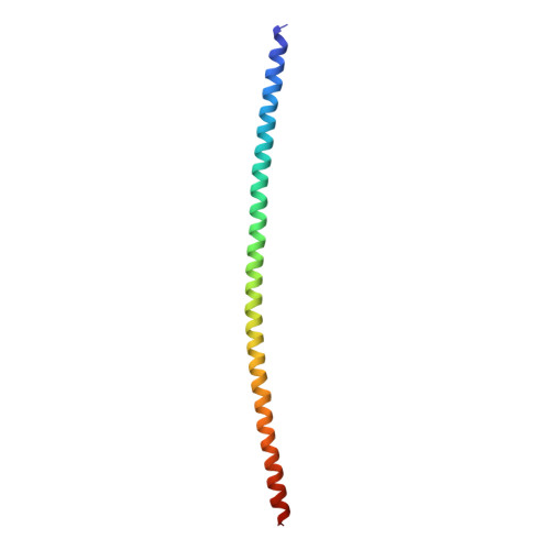
    Intermediate filaments (IFs) provide vital mechanical support in a broad array of cell types. Interference with this role causes cell fragility and accounts for a large number of human diseases. Gaining an understanding of the structure of IFs is paramount to understanding their function and designing therapeutic agents for relevant diseases. Here, we report the 2.6-Å resolution crystal structure of a complex of interacting 2B domains of keratin 5 (K5) and K14. K5 and K14 form a long-range, left-handed coiled coil, with participating α helices aligned in parallel and in register. Follow-up mutagenesis revealed that specific contacts between interacting 2B domains play a crucial role during 10-nm IF assembly, likely at the step of octamer-octamer association. The resulting structural model represents an atomic-resolution visualization of 2B-2B interactions important to filament assembly and provides insight into the defects introduced by mutations in IF genes associated with human skin diseases.


  • Organizational Affiliation

    Department of Biochemistry and Molecular Biology, Bloomberg School of Public Health, Johns Hopkins University, Baltimore, MD 21205, USA.


Macromolecules
Find similar proteins by:  (by identity cutoff)  |  3D Structure
Entity ID: 1
MoleculeChains Sequence LengthOrganismDetailsImage
Keratin, type I cytoskeletal 14
A, C
95Homo sapiensMutation(s): 1 
Gene Names: KRT14
UniProt & NIH Common Fund Data Resources
Find proteins for P02533 (Homo sapiens)
Explore P02533 
Go to UniProtKB:  P02533
PHAROS:  P02533
GTEx:  ENSG00000186847 
Entity Groups  
Sequence Clusters30% Identity50% Identity70% Identity90% Identity95% Identity100% Identity
UniProt GroupP02533
Sequence Annotations
Expand
  • Reference Sequence
Find similar proteins by:  (by identity cutoff)  |  3D Structure
Entity ID: 2
MoleculeChains Sequence LengthOrganismDetailsImage
Keratin, type II cytoskeletal 5
B, D
98Homo sapiensMutation(s): 0 
Gene Names: KRT5
UniProt & NIH Common Fund Data Resources
Find proteins for P13647 (Homo sapiens)
Explore P13647 
Go to UniProtKB:  P13647
PHAROS:  P13647
GTEx:  ENSG00000186081 
Entity Groups  
Sequence Clusters30% Identity50% Identity70% Identity90% Identity95% Identity100% Identity
UniProt GroupP13647
Sequence Annotations
Expand
  • Reference Sequence
Experimental Data & Validation

Experimental Data

  • Method: X-RAY DIFFRACTION
  • Resolution: 2.60 Å
  • R-Value Free: 0.246 
  • R-Value Work: 0.201 
  • R-Value Observed: 0.203 
  • Space Group: P 41
Unit Cell:
Length ( Å )Angle ( ˚ )
a = 56.149α = 90
b = 56.149β = 90
c = 222.054γ = 90
Software Package:
Software NamePurpose
PHENIXrefinement
HKL-2000data reduction
SCALEPACKdata scaling
PHENIXphasing

Structure Validation

View Full Validation Report


Entry History & Funding Information

Deposition Data


Funding OrganizationLocationGrant Number
National Institutes of Health/National Human Genome Research Institute (NIH/NHGRI)United States--

Revision History  (Full details and data files)

  • Version 1.0: 2020-01-22
    Type: Initial release
  • Version 1.1: 2020-04-15
    Changes: Database references, Structure summary
  • Version 1.2: 2022-03-23
    Changes: Author supporting evidence, Database references