6JFP

Crystal structure of the beta-glucosidase Bgl15


Experimental Data Snapshot

  • Method: X-RAY DIFFRACTION
  • Resolution: 2.70 Å
  • R-Value Free: 0.254 
  • R-Value Work: 0.219 
  • R-Value Observed: 0.221 

wwPDB Validation   3D Report Full Report


Ligand Structure Quality Assessment 


This is version 1.3 of the entry. See complete history


Literature

Engineering of beta-Glucosidase Bgl15 with Simultaneously Enhanced Glucose Tolerance and Thermostability To Improve Its Performance in High-Solid Cellulose Hydrolysis.

Cao, L.Chen, R.Huang, X.Li, S.Zhang, S.Yang, X.Qin, Z.Kong, W.Xie, W.Liu, Y.

(2020) J Agric Food Chem 68: 5391-5401

  • DOI: https://doi.org/10.1021/acs.jafc.0c01817
  • Primary Citation of Related Structures:  
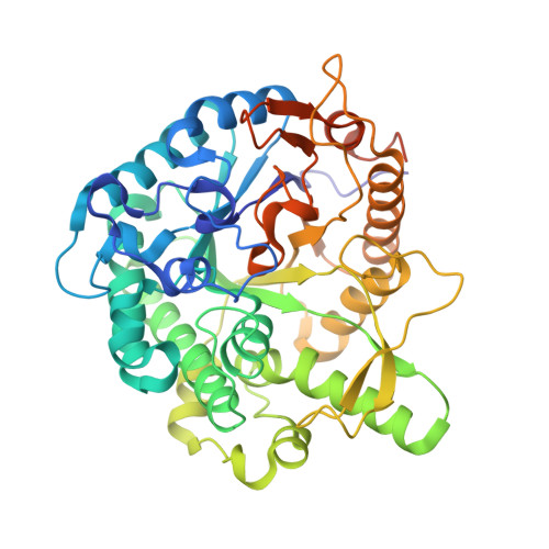
    6JFP

  • PubMed Abstract: 

    In this study, a Petri-dish-based double-layer high-throughput screening method was established to improve glucose tolerance of β-glucosidase Bgl15. Two beneficial mutations were identified, and the joint mutant 2R1 improved the half-maximal inhibitory concentration of glucose from 0.04 to 2.1 M. The crystal structure of 2R1 was subsequently determined at 2.7 Å. Structure analysis revealed that enhancement of glucose tolerance may be due to improved transglycosylation activity made possible by a hydrophobic binding site for glucose as an acceptor and more stringent control of a putative water channel. To further ameliorate the application potential of the enzyme, it was engineered to increase the half-life at 50 °C from 0.8 h (Bgl15) to 180 h (mutant 5R1). Furthermore, supplementation of 5R1 to the cellulase cocktail significantly improved glucose production from pretreated sugar cane bagasse by 38%. Consequently, this study provided an efficient approach to enhance glucose tolerance and generated a promising catalyst for cellulose saccharification.


Macromolecules
Find similar proteins by:  (by identity cutoff)  |  3D Structure
Entity ID: 1
MoleculeChains Sequence LengthOrganismDetailsImage
beta-glucosidase 15A [auth B],
B [auth A]
477uncultured bacteriumMutation(s): 0 
UniProt
Find proteins for A0A5B9BHU3 (uncultured bacterium)
Explore A0A5B9BHU3 
Go to UniProtKB:  A0A5B9BHU3
Entity Groups  
Sequence Clusters30% Identity50% Identity70% Identity90% Identity95% Identity100% Identity
UniProt GroupA0A5B9BHU3
Sequence Annotations
Expand
  • Reference Sequence
Small Molecules
Ligands 1 Unique
IDChains Name / Formula / InChI Key2D Diagram3D Interactions
BGC
Query on BGC

Download Ideal Coordinates CCD File 
C [auth A]beta-D-glucopyranose
C6 H12 O6
WQZGKKKJIJFFOK-VFUOTHLCSA-N
Experimental Data & Validation

Experimental Data

  • Method: X-RAY DIFFRACTION
  • Resolution: 2.70 Å
  • R-Value Free: 0.254 
  • R-Value Work: 0.219 
  • R-Value Observed: 0.221 
  • Space Group: P 1 21 1
Unit Cell:
Length ( Å )Angle ( ˚ )
a = 51.296α = 90
b = 93.353β = 104.2
c = 94.057γ = 90
Software Package:
Software NamePurpose
PHENIXrefinement
HKL-3000data reduction
HKL-3000data scaling
PHENIXphasing

Structure Validation

View Full Validation Report


Ligand Structure Quality Assessment 


Entry History & Funding Information

Deposition Data


Funding OrganizationLocationGrant Number
National Natural Science Foundation of ChinaChina31770075
National Natural Science Foundation of ChinaChina31870782

Revision History  (Full details and data files)

  • Version 1.0: 2020-02-12
    Type: Initial release
  • Version 1.1: 2020-07-29
    Type: Remediation
    Reason: Carbohydrate remediation
    Changes: Data collection, Derived calculations, Structure summary
  • Version 1.2: 2020-08-26
    Changes: Database references, Structure summary
  • Version 1.3: 2023-11-22
    Changes: Data collection, Database references, Refinement description