


Crystal structure of P8 from Lactobacillus rhamnosus
Cha, Y.J., Cho, H.S.To be published.
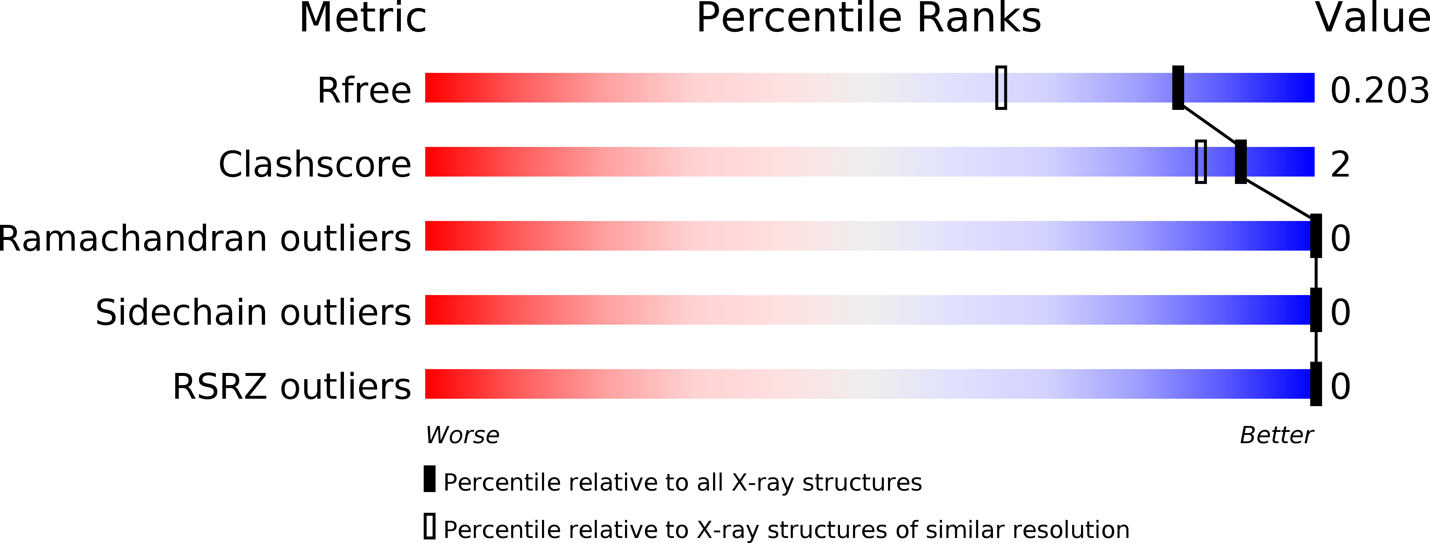
Experimental Data Snapshot
wwPDB Validation 3D Report Full Report
Entity ID: 1 | |||||
|---|---|---|---|---|---|
| Molecule | Chains | Sequence Length | Organism | Details | Image |
| P8 | 77 | Lacticaseibacillus rhamnosus | Mutation(s): 0 Gene Names: B4Q13_10920, BGK71_11245, CCE29_09795, DBP98_10520, PY66_06245, PY91_09235 | 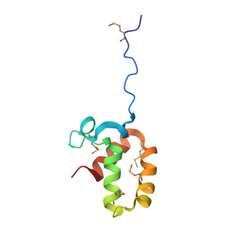 | |
UniProt | |||||
Find proteins for A0A0E3CPJ0 (Lacticaseibacillus rhamnosus) Explore A0A0E3CPJ0 Go to UniProtKB: A0A0E3CPJ0 | |||||
Entity Groups | |||||
| Sequence Clusters | 30% Identity50% Identity70% Identity90% Identity95% Identity100% Identity | ||||
| UniProt Group | A0A0E3CPJ0 | ||||
Sequence AnnotationsExpand | |||||
| |||||
| Ligands 3 Unique | |||||
|---|---|---|---|---|---|
| ID | Chains | Name / Formula / InChI Key | 2D Diagram | 3D Interactions | |
| GOL Query on GOL | H [auth A] | GLYCEROL C3 H8 O3 PEDCQBHIVMGVHV-UHFFFAOYSA-N |  | ||
| ZN Query on ZN | B [auth A], C [auth A], D [auth A], E [auth A], F [auth A] | ZINC ION Zn PTFCDOFLOPIGGS-UHFFFAOYSA-N |  | ||
| EDO Query on EDO | G [auth A] | 1,2-ETHANEDIOL C2 H6 O2 LYCAIKOWRPUZTN-UHFFFAOYSA-N |  | ||
| Modified Residues 1 Unique | |||||
|---|---|---|---|---|---|
| ID | Chains | Type | Formula | 2D Diagram | Parent |
| MSE Query on MSE | A | L-PEPTIDE LINKING | C5 H11 N O2 Se |  | MET |
| Length ( Å ) | Angle ( ˚ ) |
|---|---|
| a = 40.86 | α = 90 |
| b = 40.86 | β = 90 |
| c = 107.01 | γ = 90 |
| Software Name | Purpose |
|---|---|
| REFMAC | refinement |
| MOSFLM | data reduction |
| Aimless | data scaling |
| AutoSol | phasing |
RCSB PDB (citation) is hosted by
RCSB PDB is a member of the

