6I9Y

The 2.14 A X-ray crystal structure of Sporosarcina pasteurii urease in complex with Au(I) ions


Experimental Data Snapshot

  • Method: X-RAY DIFFRACTION
  • Resolution: 2.14 Å
  • R-Value Free: 0.208 
  • R-Value Work: 0.171 
  • R-Value Observed: 0.173 

wwPDB Validation   3D Report Full Report


This is version 1.1 of the entry. See complete history


Literature

Inhibition Mechanism of Urease by Au(III) Compounds Unveiled by X-ray Diffraction Analysis.

Mazzei, L.Wenzel, M.N.Cianci, M.Palombo, M.Casini, A.Ciurli, S.

(2019) ACS Med Chem Lett 10: 564-570

  • DOI: https://doi.org/10.1021/acsmedchemlett.8b00585
  • Primary Citation of Related Structures:  
    6I9Y

  • PubMed Abstract: 

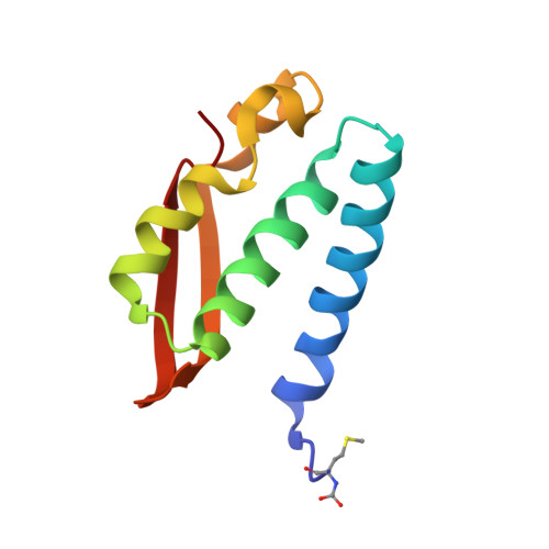
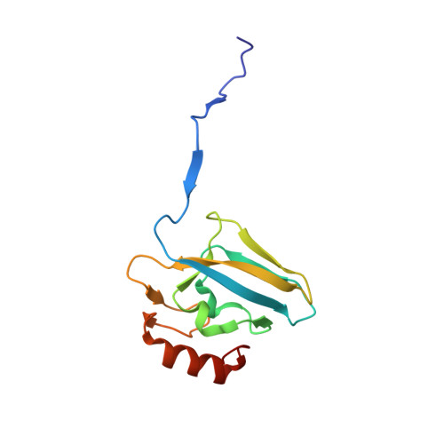
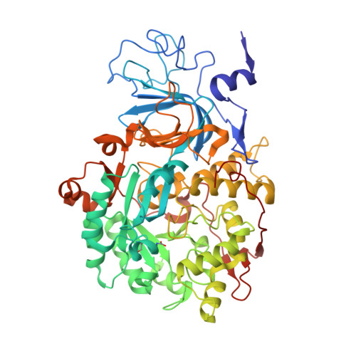
    The nickel-dependent enzyme urease is a virulence factor for a large number of critical human pathogens, making this enzyme a potential target of therapeutics for the treatment of resistant bacterial infections. In the search for novel urease inhibitors, five selected coordination and organometallic Au(III) compounds containing N N or C N and C N N ligands were tested for their inhibitory effects against Canavalia ensiformis (jack bean) urease. The results showed potent inhibition effects with IC 50 values in the nanomolar range. The 2.14 Å resolution crystal structure of Sporosarcina pasteurii urease inhibited by the most effective Au(III) compound [Au(PbImMe)Cl 2 ]PF 6 (PbImMe = 1-methyl-2-(pyridin-2-yl)-benzimidazole) reveals the presence of two Au ions bound to the conserved triad αCys322/αHis323/αMet367. The binding of the Au ions to these residues blocks the movement of a flap, located at the edge of the active site channel and essential for enzyme catalysis, completely obliterating the catalytic activity of urease. Overall, the obtained results constitute the basis for the design of new gold complexes as selective urease inhibitors with future antibacterial applications.


  • Organizational Affiliation

    Laboratory of Bioinorganic Chemistry, Department of Pharmacy and Biotechnology, University of Bologna, Viale Giuseppe Fanin 40, I-40127 Bologna, Italy.


Macromolecules
Find similar proteins by:  (by identity cutoff)  |  3D Structure
Entity ID: 1
MoleculeChains Sequence LengthOrganismDetailsImage
Urease subunit gamma100Sporosarcina pasteuriiMutation(s): 0 
EC: 3.5.1.5
UniProt
Find proteins for A0A0H3YGY5 (Sporosarcina pasteurii)
Explore A0A0H3YGY5 
Go to UniProtKB:  A0A0H3YGY5
Entity Groups  
Sequence Clusters30% Identity50% Identity70% Identity90% Identity95% Identity100% Identity
UniProt GroupA0A0H3YGY5
Sequence Annotations
Expand
  • Reference Sequence
Find similar proteins by:  (by identity cutoff)  |  3D Structure
Entity ID: 2
MoleculeChains Sequence LengthOrganismDetailsImage
Urease subunit beta122Sporosarcina pasteuriiMutation(s): 0 
EC: 3.5.1.5
UniProt
Find proteins for P41021 (Sporosarcina pasteurii)
Explore P41021 
Go to UniProtKB:  P41021
Entity Groups  
Sequence Clusters30% Identity50% Identity70% Identity90% Identity95% Identity100% Identity
UniProt GroupP41021
Sequence Annotations
Expand
  • Reference Sequence
Find similar proteins by:  (by identity cutoff)  |  3D Structure
Entity ID: 3
MoleculeChains Sequence LengthOrganismDetailsImage
Urease subunit alpha570Sporosarcina pasteuriiMutation(s): 0 
EC: 3.5.1.5
UniProt
Find proteins for P41020 (Sporosarcina pasteurii)
Explore P41020 
Go to UniProtKB:  P41020
Entity Groups  
Sequence Clusters30% Identity50% Identity70% Identity90% Identity95% Identity100% Identity
UniProt GroupP41020
Sequence Annotations
Expand
  • Reference Sequence
Small Molecules
Ligands 5 Unique
IDChains Name / Formula / InChI Key2D Diagram3D Interactions
AU
Query on AU

Download Ideal Coordinates CCD File 
V [auth C],
W [auth C],
X [auth C]
GOLD ION
Au
ZBKIUFWVEIBQRT-UHFFFAOYSA-N
SO4
Query on SO4

Download Ideal Coordinates CCD File 
E [auth A]
F [auth A]
I [auth B]
J [auth B]
T [auth C]
E [auth A],
F [auth A],
I [auth B],
J [auth B],
T [auth C],
U [auth C],
Y [auth C]
SULFATE ION
O4 S
QAOWNCQODCNURD-UHFFFAOYSA-L
EDO
Query on EDO

Download Ideal Coordinates CCD File 
AA [auth C]
D [auth A]
G [auth B]
H [auth B]
N [auth C]
AA [auth C],
D [auth A],
G [auth B],
H [auth B],
N [auth C],
O [auth C],
P [auth C],
Q [auth C],
R [auth C],
S [auth C],
Z [auth C]
1,2-ETHANEDIOL
C2 H6 O2
LYCAIKOWRPUZTN-UHFFFAOYSA-N
NI
Query on NI

Download Ideal Coordinates CCD File 
K [auth C],
L [auth C]
NICKEL (II) ION
Ni
VEQPNABPJHWNSG-UHFFFAOYSA-N
OH
Query on OH

Download Ideal Coordinates CCD File 
M [auth C]HYDROXIDE ION
H O
XLYOFNOQVPJJNP-UHFFFAOYSA-M
Modified Residues  2 Unique
IDChains TypeFormula2D DiagramParent
CXM
Query on CXM
A
L-PEPTIDE LINKINGC6 H11 N O4 SMET
KCX
Query on KCX
C
L-PEPTIDE LINKINGC7 H14 N2 O4LYS
Experimental Data & Validation

Experimental Data

  • Method: X-RAY DIFFRACTION
  • Resolution: 2.14 Å
  • R-Value Free: 0.208 
  • R-Value Work: 0.171 
  • R-Value Observed: 0.173 
  • Space Group: P 63 2 2
Unit Cell:
Length ( Å )Angle ( ˚ )
a = 132.291α = 90
b = 132.291β = 90
c = 190.435γ = 120
Software Package:
Software NamePurpose
REFMACrefinement
XDSdata reduction
Aimlessdata scaling
REFMACphasing

Structure Validation

View Full Validation Report


Entry History 

Deposition Data

Revision History  (Full details and data files)

  • Version 1.0: 2019-05-01
    Type: Initial release
  • Version 1.1: 2024-01-24
    Changes: Data collection, Database references, Derived calculations, Refinement description