The Extreme Structural Plasticity in the CYP153 Subfamily of P450s Directs Development of Designer Hydroxylases.
Fiorentini, F., Hatzl, A.M., Schmidt, S., Savino, S., Glieder, A., Mattevi, A.(2018) Biochemistry 57: 6701-6714
- PubMed: 30398864
- DOI: https://doi.org/10.1021/acs.biochem.8b01052
- Primary Citation of Related Structures:
6HQD, 6HQG, 6HQW - PubMed Abstract:
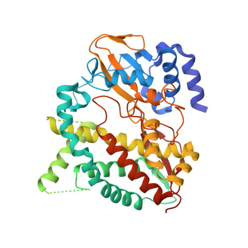
CYP153s are bacterial class I P450 enzymes traditionally described as alkane hydroxylases with a high terminal regioselectivity. They have been more recently shown to also catalyze hydroxylations at nonactivated carbon atoms of small heterocycles. The aim of our work was to perform an extensive characterization of this subfamily in order to deliver a toolbox of CYP153 enzymes for further development as biocatalysts. Through the screening of recently sequenced bacterial genomes, 20 CYP153s were selected, comprising 17 single monooxygenase domains and three multidomain variants, where the monooxygenase domain is naturally fused to its redox partners in a single polypeptide chain. The 20 novel variants were heterologously expressed, and their activity was screened toward octane and small heterocycles. A more extended substrate characterization was then performed on three representative candidates, and their crystal structures were unveiled and compared with those of the known CYP153A7 and CYP153A33. The tested enzymes displayed a wide range of activities, ranging from Ω and Ω-1 hydroxylations of lauric acid to indigo-generating indole modification. The comparative analysis highlighted a conserved architecture and amino acid composition of the catalytic core close to the heme, while showing a huge degree of structural plasticity and flexibility in those regions hosting the substrate recognition sites. Although dealing with this type of conformational variability adds a layer of complexity and difficulty to structure-based protein engineering, such diversity in substrate acceptance and recognition promotes the investigated CYP153s as a prime choice for tailoring designer hydroxylases.
Organizational Affiliation:
Department of Biology and Biotechnology , University of Pavia , via Ferrata 9 , Pavia 27100 , Italy.