


High Resolution Structures of Viral Neuraminidase with Drugs Bound in the Active Site. (In preparation)
Salinger, M.T., Hobbs, J.R., Murray, J.W., Laver, W.G., Kuhn, P., Garman, E.F.To be published.
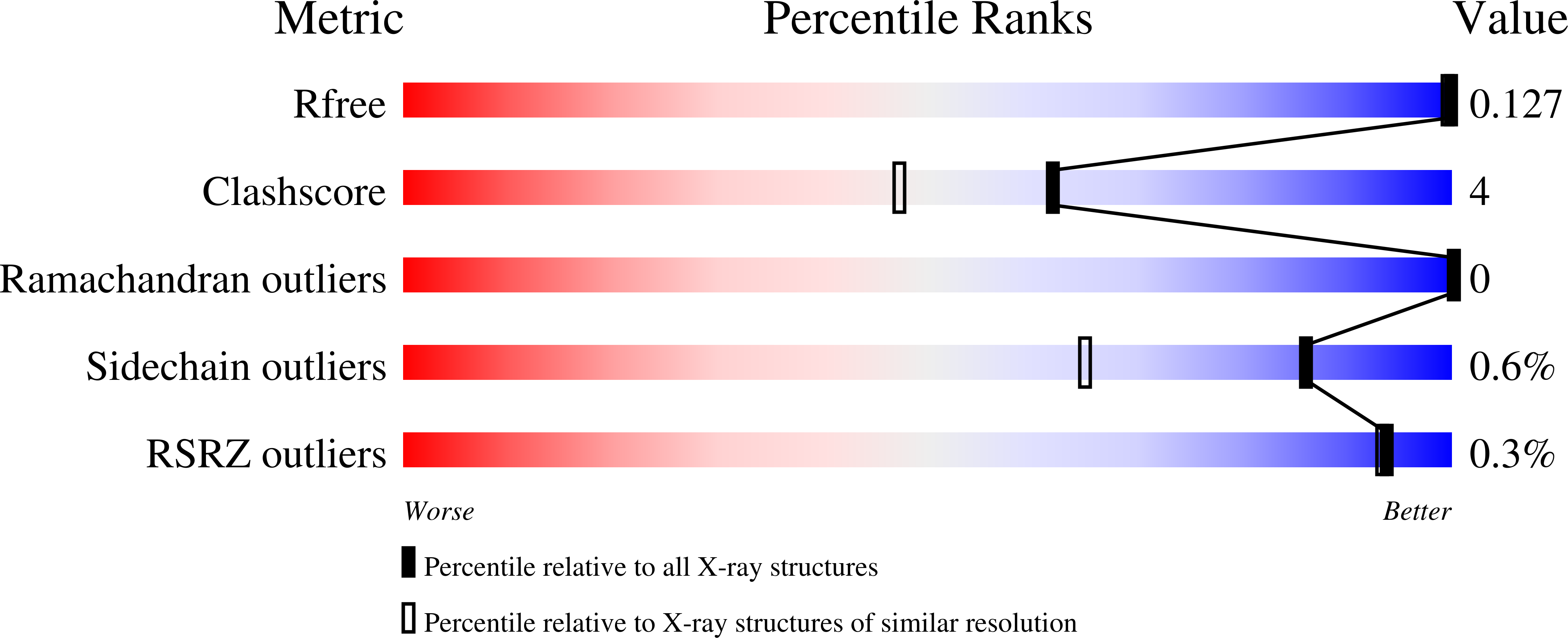
Experimental Data Snapshot
Entity ID: 1 | |||||
|---|---|---|---|---|---|
| Molecule | Chains | Sequence Length | Organism | Details | Image |
| Neuraminidase | 388 | Influenza A virus (A/tern/Australia/G70C/1975(H11N9)) | Mutation(s): 0 Gene Names: NA EC: 3.2.1.18 | 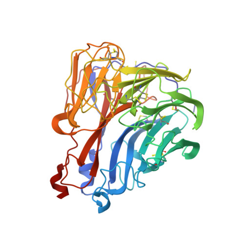 | |
UniProt | |||||
Find proteins for P03472 (Influenza A virus (strain A/Tern/Australia/G70C/1975 H11N9)) Explore P03472 Go to UniProtKB: P03472 | |||||
Entity Groups | |||||
| Sequence Clusters | 30% Identity50% Identity70% Identity90% Identity95% Identity100% Identity | ||||
| UniProt Group | P03472 | ||||
Sequence AnnotationsExpand | |||||
| |||||
Entity ID: 2 | |||||
|---|---|---|---|---|---|
| Molecule | Chains | Length | 2D Diagram | Glycosylation | 3D Interactions |
| alpha-D-mannopyranose-(1-2)-alpha-D-mannopyranose-(1-2)-alpha-D-mannopyranose-(1-3)-[alpha-D-mannopyranose-(1-3)-[alpha-D-mannopyranose-(1-6)]alpha-D-mannopyranose-(1-6)]beta-D-mannopyranose-(1-4)-2-acetamido-2-deoxy-beta-D-glucopyranose-(1-4)-2-acetamido-2-deoxy-beta-D-glucopyranose | B | 9 |  | N-Glycosylation | |
Glycosylation Resources | |||||
GlyTouCan: G68668TB GlyCosmos: G68668TB GlyGen: G68668TB | |||||
| Ligands 4 Unique | |||||
|---|---|---|---|---|---|
| ID | Chains | Name / Formula / InChI Key | 2D Diagram | 3D Interactions | |
| ZMR Query on ZMR | J [auth A] | ZANAMIVIR C12 H20 N4 O7 ARAIBEBZBOPLMB-UFGQHTETSA-N |  | ||
| GOL Query on GOL | G [auth A], H [auth A], I [auth A] | GLYCEROL C3 H8 O3 PEDCQBHIVMGVHV-UHFFFAOYSA-N |  | ||
| CA Query on CA | E [auth A], F [auth A] | CALCIUM ION Ca BHPQYMZQTOCNFJ-UHFFFAOYSA-N |  | ||
| K Query on K | K [auth A] | POTASSIUM ION K NPYPAHLBTDXSSS-UHFFFAOYSA-N |  | ||
| Length ( Å ) | Angle ( ˚ ) |
|---|---|
| a = 180.935 | α = 90 |
| b = 180.935 | β = 90 |
| c = 180.935 | γ = 90 |
| Software Name | Purpose |
|---|---|
| REFMAC | refinement |
| MOSFLM | data reduction |
| SCALA | data scaling |
| REFMAC | phasing |
| Funding Organization | Location | Grant Number |
|---|---|---|
| United Kingdom | -- |
RCSB PDB (citation) is hosted by
RCSB PDB is a member of the


