6HCS

Crystal structure of CaM-peptide complex containing AzF at position 108


Experimental Data Snapshot

  • Method: X-RAY DIFFRACTION
  • Resolution: 2.00 Å
  • R-Value Free: 0.294 
  • R-Value Work: 0.260 
  • R-Value Observed: 0.262 

wwPDB Validation   3D Report Full Report


This is version 1.2 of the entry. See complete history


Literature

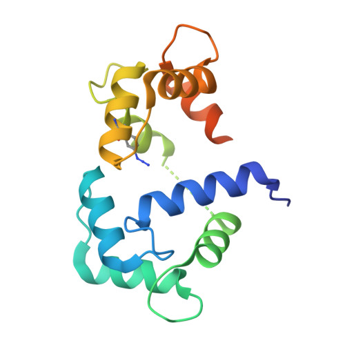
Conformation-specific detection of calmodulin binding using the unnatural amino acid p-azido-phenylalanine (AzF) as an IR-sensor.

Creon, A.Josts, I.Niebling, S.Huse, N.Tidow, H.

(2018) Struct Dyn 5: 064701-064701

  • DOI: https://doi.org/10.1063/1.5053466
  • Primary Citation of Related Structures:  
    6HCS

  • PubMed Abstract: 

    Calmodulin (CaM) is a very conserved, ubiquitous, eukaryotic protein that binds four Ca 2+ ions with high affinity. It acts as a calcium sensor by translating Ca 2+ signals into cellular processes such as metabolism, inflammation, immune response, memory, and muscle contraction. Calcium binding to CaM leads to conformational changes that enable Ca 2+ /CaM to recognize and bind various target proteins with high affinity. The binding mode and binding partners of CaM are very diverse, and a consensus binding sequence is lacking. Here, we describe an elegant system that allows conformation-specific detection of CaM-binding to its binding partners. We incorporate the unnatural amino acid p-azido-phenylalanine (AzF) in different positions of CaM and follow its unique spectral signature by infrared (IR)-spectroscopy of the azido stretching vibration. Our results suggest that the AzF vibrational probe is sensitive to the chemical environment in different CaM/CaM-binding domain (CaMBD) complexes, which allows differentiating between different binding motifs according to the spectral characteristics of the azido stretching mode. We corroborate our results with a crystal structure of AzF-labelled CaM (CaM108AzF) in complex with a binding peptide from calmodulin-dependent protein kinase IIα identifying the structural basis for the observed IR frequency shifts.


Macromolecules
Find similar proteins by:  (by identity cutoff)  |  3D Structure
Entity ID: 1
MoleculeChains Sequence LengthOrganismDetailsImage
Calmodulin-1
A, C, E, G
167Homo sapiensMutation(s): 1 
Gene Names: CALM1CALMCAMCAM1
UniProt & NIH Common Fund Data Resources
Find proteins for P0DP23 (Homo sapiens)
Explore P0DP23 
Go to UniProtKB:  P0DP23
PHAROS:  P0DP23
GTEx:  ENSG00000198668 
Entity Groups  
Sequence Clusters30% Identity50% Identity70% Identity90% Identity95% Identity100% Identity
UniProt GroupP0DP23
Sequence Annotations
Expand
  • Reference Sequence

Find similar proteins by:  Sequence   |   3D Structure  

Entity ID: 2
MoleculeChains Sequence LengthOrganismDetailsImage
Calcium/calmodulin-dependent protein kinase type II subunit beta
B, D, F, H
25Rattus norvegicusMutation(s): 0 
EC: 2.7.11.17
UniProt
Find proteins for P08413 (Rattus norvegicus)
Explore P08413 
Go to UniProtKB:  P08413
Entity Groups  
Sequence Clusters30% Identity50% Identity70% Identity90% Identity95% Identity100% Identity
UniProt GroupP08413
Sequence Annotations
Expand
  • Reference Sequence
Small Molecules
Ligands 1 Unique
IDChains Name / Formula / InChI Key2D Diagram3D Interactions
CA
Query on CA

Download Ideal Coordinates CCD File 
I [auth A]
J [auth A]
K [auth A]
L [auth A]
M [auth C]
I [auth A],
J [auth A],
K [auth A],
L [auth A],
M [auth C],
N [auth C],
O [auth C],
P [auth C],
Q [auth E],
R [auth E],
S [auth E],
T [auth E],
U [auth G],
V [auth G],
W [auth G],
X [auth G]
CALCIUM ION
Ca
BHPQYMZQTOCNFJ-UHFFFAOYSA-N
Modified Residues  1 Unique
IDChains TypeFormula2D DiagramParent
4II
Query on 4II
A, C, E, G
L-PEPTIDE LINKINGC9 H10 N4 O2PHE
Experimental Data & Validation

Experimental Data

  • Method: X-RAY DIFFRACTION
  • Resolution: 2.00 Å
  • R-Value Free: 0.294 
  • R-Value Work: 0.260 
  • R-Value Observed: 0.262 
  • Space Group: P 1 21 1
Unit Cell:
Length ( Å )Angle ( ˚ )
a = 76.263α = 90
b = 37.214β = 100.21
c = 121.193γ = 90
Software Package:
Software NamePurpose
PHENIXrefinement
XDSdata reduction
Aimlessdata scaling
PHASERphasing

Structure Validation

View Full Validation Report


Entry History 

Deposition Data

Revision History  (Full details and data files)

  • Version 1.0: 2018-12-05
    Type: Initial release
  • Version 1.1: 2018-12-19
    Changes: Data collection, Derived calculations
  • Version 1.2: 2024-01-17
    Changes: Data collection, Database references, Derived calculations, Refinement description