6H16

Structure of LRP6 P3E3P4E4 in complex with VHH L-P2-D07


Experimental Data Snapshot

  • Method: X-RAY DIFFRACTION
  • Resolution: 2.90 Å
  • R-Value Free: 0.252 
  • R-Value Work: 0.211 
  • R-Value Observed: 0.213 

wwPDB Validation   3D Report Full Report


Ligand Structure Quality Assessment 


This is version 2.1 of the entry. See complete history


Literature

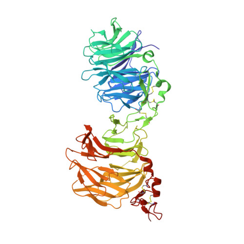
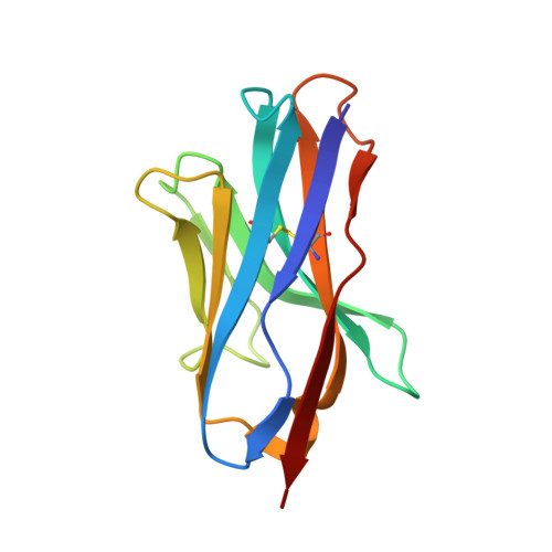
Anti-LRP5/6 VHHs promote differentiation of Wnt-hypersensitive intestinal stem cells.

Fenderico, N.van Scherpenzeel, R.C.Goldflam, M.Proverbio, D.Jordens, I.Kralj, T.Stryeck, S.Bass, T.Z.Hermans, G.Ullman, C.Aastrup, T.Gros, P.Maurice, M.M.

(2019) Nat Commun 10: 365-365

  • DOI: https://doi.org/10.1038/s41467-018-08172-z
  • Primary Citation of Related Structures:  
    6H15, 6H16

  • PubMed Abstract: 

    Wnt-induced β-catenin-mediated transcription is a driving force for stem cell self-renewal during adult tissue homeostasis. Enhanced Wnt receptor expression due to mutational inactivation of the ubiquitin ligases RNF43/ZNRF3 recently emerged as a leading cause for cancer development. Consequently, targeting canonical Wnt receptors such as LRP5/6 holds great promise for treatment of such cancer subsets. Here, we employ CIS display technology to identify single-domain antibody fragments (VHH) that bind the LRP6 P3E3P4E4 region with nanomolar affinity and strongly inhibit Wnt3/3a-induced β-catenin-mediated transcription in cells, while leaving Wnt1 responses unaffected. Structural analysis reveal that individual VHHs variably employ divergent antigen-binding regions to bind a similar surface in the third β-propeller of LRP5/6, sterically interfering with Wnt3/3a binding. Importantly, anti-LRP5/6 VHHs block the growth of Wnt-hypersensitive Rnf43/Znrf3-mutant intestinal organoids through stem cell exhaustion and collective terminal differentiation. Thus, VHH-mediated targeting of LRP5/6 provides a promising differentiation-inducing strategy for treatment of Wnt-hypersensitive tumors.


  • Organizational Affiliation

    Oncode Institute and Department of Cell Biology, Center for Molecular Medicine, University Medical Center Utrecht, 3584 CX, Utrecht, The Netherlands.


Macromolecules
Find similar proteins by:  (by identity cutoff)  |  3D Structure
Entity ID: 1
MoleculeChains Sequence LengthOrganismDetailsImage
Low-density lipoprotein receptor-related protein 6618Homo sapiensMutation(s): 0 
Gene Names: LRP6
UniProt & NIH Common Fund Data Resources
Find proteins for O75581 (Homo sapiens)
Explore O75581 
Go to UniProtKB:  O75581
PHAROS:  O75581
GTEx:  ENSG00000070018 
Entity Groups  
Sequence Clusters30% Identity50% Identity70% Identity90% Identity95% Identity100% Identity
UniProt GroupO75581
Sequence Annotations
Expand
  • Reference Sequence
Find similar proteins by:  (by identity cutoff)  |  3D Structure
Entity ID: 2
MoleculeChains Sequence LengthOrganismDetailsImage
VHH L-P2-D07119Lama glamaMutation(s): 0 
Entity Groups  
Sequence Clusters30% Identity50% Identity70% Identity90% Identity95% Identity100% Identity
Sequence Annotations
Expand
  • Reference Sequence
Oligosaccharides

Help

Entity ID: 3
MoleculeChains Length2D Diagram Glycosylation3D Interactions
beta-D-mannopyranose-(1-4)-2-acetamido-2-deoxy-beta-D-glucopyranose-(1-4)-2-acetamido-2-deoxy-beta-D-glucopyranose
C, D, E
3N-Glycosylation
Glycosylation Resources
GlyTouCan:  G15407YE
GlyCosmos:  G15407YE
GlyGen:  G15407YE
Entity ID: 4
MoleculeChains Length2D Diagram Glycosylation3D Interactions
alpha-D-mannopyranose-(1-3)-beta-D-mannopyranose-(1-4)-2-acetamido-2-deoxy-beta-D-glucopyranose-(1-4)-2-acetamido-2-deoxy-beta-D-glucopyranose
F
4N-Glycosylation
Glycosylation Resources
GlyTouCan:  G81315DD
GlyCosmos:  G81315DD
GlyGen:  G81315DD
Small Molecules
Ligands 2 Unique
IDChains Name / Formula / InChI Key2D Diagram3D Interactions
NAG
Query on NAG

Download Ideal Coordinates CCD File 
G [auth A]2-acetamido-2-deoxy-beta-D-glucopyranose
C8 H15 N O6
OVRNDRQMDRJTHS-FMDGEEDCSA-N
CA
Query on CA

Download Ideal Coordinates CCD File 
H [auth A]CALCIUM ION
Ca
BHPQYMZQTOCNFJ-UHFFFAOYSA-N
Experimental Data & Validation

Experimental Data

  • Method: X-RAY DIFFRACTION
  • Resolution: 2.90 Å
  • R-Value Free: 0.252 
  • R-Value Work: 0.211 
  • R-Value Observed: 0.213 
  • Space Group: P 21 21 21
Unit Cell:
Length ( Å )Angle ( ˚ )
a = 92.795α = 90
b = 105.954β = 90
c = 164.191γ = 90
Software Package:
Software NamePurpose
PHENIXrefinement
Aimlessdata scaling
PDB_EXTRACTdata extraction
DIALSdata reduction
PHASERphasing

Structure Validation

View Full Validation Report


Ligand Structure Quality Assessment 


Entry History & Funding Information

Deposition Data


Funding OrganizationLocationGrant Number
Netherlands Organisation for Scientific ResearchNetherlands01.80.104.00

Revision History  (Full details and data files)

  • Version 1.0: 2019-01-30
    Type: Initial release
  • Version 2.0: 2020-07-29
    Type: Remediation
    Reason: Carbohydrate remediation
    Changes: Atomic model, Data collection, Derived calculations, Structure summary
  • Version 2.1: 2024-01-17
    Changes: Data collection, Database references, Derived calculations, Refinement description, Structure summary