Neutron and X-ray crystal structures of Lactobacillus brevis alcohol dehydrogenase reveal new insights into hydrogen-bonding pathways.
Hermann, J., Nowotny, P., Schrader, T.E., Biggel, P., Hekmat, D., Weuster-Botz, D.(2018) Acta Crystallogr F Struct Biol Commun 74: 754-764
- PubMed: 30511668
- DOI: https://doi.org/10.1107/S2053230X18015273
- Primary Citation of Related Structures:
6H07, 6H1M - PubMed Abstract:
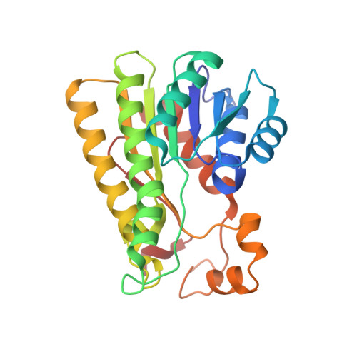
Lactobacillus brevis alcohol dehydrogenase (LbADH) is a well studied homotetrameric enzyme which catalyzes the enantioselective reduction of prochiral ketones to the corresponding secondary alcohols. LbADH is stable and enzymatically active at elevated temperatures and accepts a broad range of substrates, making it a valuable tool in industrial biocatalysis. Here, the expression, purification and crystallization of LbADH to generate large, single crystals with a volume of up to 1 mm 3 suitable for neutron diffraction studies are described. Neutron diffraction data were collected from an H/D-exchanged LbADH crystal using the BIODIFF instrument at the Heinz Maier-Leibnitz Zentrum (MLZ), Garching, Germany to a resolution d min of 2.15 Å in 16 days. This allowed the first neutron crystal structure of LbADH to be determined. The neutron structure revealed new details of the hydrogen-bonding network originating from the ion-binding site of LbADH and provided new insights into the reasons why divalent magnesium (Mg 2+ ) or manganese (Mn 2+ ) ions are necessary for its activity. X-ray diffraction data were obtained from the same crystal at the European Synchrotron Radiation Facility (ESRF), Grenoble, France to a resolution d min of 1.48 Å. The high-resolution X-ray structure suggested partial occupancy of Mn 2+ and Mg 2+ at the ion-binding site. This is supported by the different binding affinity of Mn 2+ and Mg 2+ to the tetrameric structure calculated via free-energy molecular-dynamics simulations.
Organizational Affiliation:
Institute of Biochemical Engineering, Technical University of Munich, Boltzmannstrasse 15, 85748 Garching, Germany.