6GUO

Siderophore hydrolase EstA from Aspergillus nidulans


Experimental Data Snapshot

  • Method: X-RAY DIFFRACTION
  • Resolution: 1.75 Å
  • R-Value Free: 0.203 
  • R-Value Work: 0.163 
  • R-Value Observed: 0.165 

wwPDB Validation   3D Report Full Report


This is version 1.2 of the entry. See complete history


Literature

Iron Scavenging in Aspergillus Species: Structural and Biochemical Insights into Fungal Siderophore Esterases.

Ecker, F.Haas, H.Groll, M.Huber, E.M.

(2018) Angew Chem Int Ed Engl 57: 14624-14629

  • DOI: https://doi.org/10.1002/anie.201807093
  • Primary Citation of Related Structures:  
    6GUD, 6GUG, 6GUI, 6GUL, 6GUN, 6GUO, 6GUP, 6GUR

  • PubMed Abstract: 

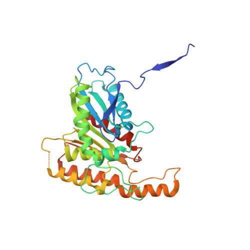
    Fungi utilize high-affinity chelators termed siderophores with chemically diverse structures to scavenge the essential nutrient iron from their surroundings. Since they are among the strongest known Fe 3+ binding agents, intracellular release of the heavy metal atom is facilitated by the activity of specific hydrolases. In this work, we report the characterization and X-ray crystal structures of four siderophore esterases: AfEstB and AfSidJ from Aspergillus fumigatus, as well as AnEstB and AnEstA from Aspergillus nidulans. Even though they all display the conserved α/β-hydrolase fold, we found significant structural and enzymatic discrepancies in their adaption to both related and chemically diverse substrates. A structure of AfEstB in complex with its substrate triacetylfusarinine C gives insight into the active enzyme and shows tetrahedral coordination between the catalytic serine and the scissile ester bond.


  • Organizational Affiliation

    Center for Integrated Protein Science Munich (CIPSM), Department of Chemistry, Technische Universität München, Lichtenbergstraße 4, 85748, Garching, Germany.


Macromolecules
Find similar proteins by:  (by identity cutoff)  |  3D Structure
Entity ID: 1
MoleculeChains Sequence LengthOrganismDetailsImage
Putative siderophore-degrading esterase (Eurofung)
A, B, C, D
304Aspergillus nidulansMutation(s): 0 
Gene Names: ANIA_07801
UniProt
Find proteins for Q5AV79 (Emericella nidulans (strain FGSC A4 / ATCC 38163 / CBS 112.46 / NRRL 194 / M139))
Explore Q5AV79 
Go to UniProtKB:  Q5AV79
Entity Groups  
Sequence Clusters30% Identity50% Identity70% Identity90% Identity95% Identity100% Identity
UniProt GroupQ5AV79
Sequence Annotations
Expand
  • Reference Sequence
Small Molecules
Ligands 3 Unique
IDChains Name / Formula / InChI Key2D Diagram3D Interactions
PGE
Query on PGE

Download Ideal Coordinates CCD File 
E [auth A]
F [auth A]
I [auth B]
J [auth B]
M [auth C]
E [auth A],
F [auth A],
I [auth B],
J [auth B],
M [auth C],
P [auth D],
Q [auth D]
TRIETHYLENE GLYCOL
C6 H14 O4
ZIBGPFATKBEMQZ-UHFFFAOYSA-N
GOL
Query on GOL

Download Ideal Coordinates CCD File 
N [auth C]GLYCEROL
C3 H8 O3
PEDCQBHIVMGVHV-UHFFFAOYSA-N
CL
Query on CL

Download Ideal Coordinates CCD File 
G [auth A]
H [auth A]
K [auth B]
L [auth B]
O [auth C]
G [auth A],
H [auth A],
K [auth B],
L [auth B],
O [auth C],
R [auth D]
CHLORIDE ION
Cl
VEXZGXHMUGYJMC-UHFFFAOYSA-M
Experimental Data & Validation

Experimental Data

  • Method: X-RAY DIFFRACTION
  • Resolution: 1.75 Å
  • R-Value Free: 0.203 
  • R-Value Work: 0.163 
  • R-Value Observed: 0.165 
  • Space Group: P 21 21 2
Unit Cell:
Length ( Å )Angle ( ˚ )
a = 105.57α = 90
b = 236.19β = 90
c = 51.61γ = 90
Software Package:
Software NamePurpose
REFMACrefinement
XDSdata reduction
XSCALEdata scaling
PHASERphasing

Structure Validation

View Full Validation Report


Entry History & Funding Information

Deposition Data


Funding OrganizationLocationGrant Number
German Research FoundationGermanyGR 1861/8-1
Austrian Science FundAustriaFWF I 1346-B21

Revision History  (Full details and data files)

  • Version 1.0: 2018-08-15
    Type: Initial release
  • Version 1.1: 2018-10-31
    Changes: Data collection, Database references
  • Version 1.2: 2024-01-17
    Changes: Data collection, Database references, Refinement description