


hPDE4D2 structure with inhibitor NPD-1086
Salado, I.G., Moreno, C., Sakaine, G., Singh, A.K., Blaazer, A.R., Siderius, M., Matheeussen, A., Gul, S., Maes, L., Leurs, R., Brown, D.G., Augustyns, K.To be published.
Experimental Data Snapshot
Entity ID: 1 | |||||
|---|---|---|---|---|---|
| Molecule | Chains | Sequence Length | Organism | Details | Image |
| cAMP-specific 3',5'-cyclic phosphodiesterase 4D | 364 | Homo sapiens | Mutation(s): 0 Gene Names: PDE4D, DPDE3 EC: 3.1.4.53 | 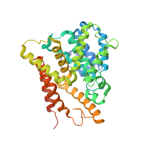 | |
UniProt & NIH Common Fund Data Resources | |||||
Find proteins for Q08499 (Homo sapiens) Explore Q08499 Go to UniProtKB: Q08499 | |||||
PHAROS: Q08499 GTEx: ENSG00000113448 | |||||
Entity Groups | |||||
| Sequence Clusters | 30% Identity50% Identity70% Identity90% Identity95% Identity100% Identity | ||||
| UniProt Group | Q08499 | ||||
Sequence AnnotationsExpand | |||||
| |||||
| Ligands 5 Unique | |||||
|---|---|---|---|---|---|
| ID | Chains | Name / Formula / InChI Key | 2D Diagram | 3D Interactions | |
| D5Z (Subject of Investigation/LOI) Query on D5Z | M [auth A], N [auth A], Z [auth B] | (4aS,8aR)-2-[1-(2-aminoquinazolin-4-yl)piperidin-4-yl]-4-(3,4-dimethoxyphenyl)-1,2,4a,5,8,8a-hexahydrophthalazin-1-one C29 H32 N6 O3 SEVYDVWEYJOUSZ-LEWJYISDSA-N |  | ||
| EPE Query on EPE | CA [auth B], Q [auth A] | 4-(2-HYDROXYETHYL)-1-PIPERAZINE ETHANESULFONIC ACID C8 H18 N2 O4 S JKMHFZQWWAIEOD-UHFFFAOYSA-N |  | ||
| ZN Query on ZN | BA [auth B], P [auth A] | ZINC ION Zn PTFCDOFLOPIGGS-UHFFFAOYSA-N |  | ||
| EDO Query on EDO | C [auth A] D [auth A] DA [auth B] E [auth A] F [auth A] | 1,2-ETHANEDIOL C2 H6 O2 LYCAIKOWRPUZTN-UHFFFAOYSA-N |  | ||
| MG Query on MG | AA [auth B], O [auth A] | MAGNESIUM ION Mg JLVVSXFLKOJNIY-UHFFFAOYSA-N |  | ||
| Length ( Å ) | Angle ( ˚ ) |
|---|---|
| a = 95.068 | α = 90 |
| b = 158.233 | β = 90 |
| c = 111.405 | γ = 90 |
| Software Name | Purpose |
|---|---|
| REFMAC | refinement |
| XDS | data reduction |
| autoPROC | data scaling |
| PHASER | phasing |
RCSB PDB (citation) is hosted by
RCSB PDB is a member of the

