


TbrPDEB1 structure with inhibitor NPD-356
Salado, I.G., Moreno, C., Sakaine, G., Singh, A.K., Blaazer, A.R., Siderius, M., Matheeussen, A., Gul, S., Maes, L., Leurs, R., Brown, D.G., Augustyns, K.To be published.
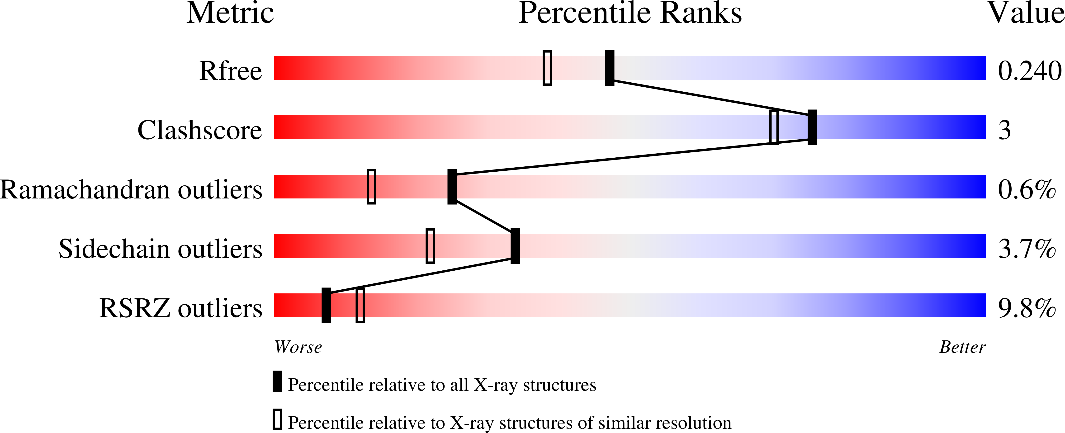
Experimental Data Snapshot
Entity ID: 1 | |||||
|---|---|---|---|---|---|
| Molecule | Chains | Sequence Length | Organism | Details | Image |
| Phosphodiesterase | 360 | Trypanosoma brucei | Mutation(s): 0 Gene Names: PDEB1 EC: 3.1.4 | 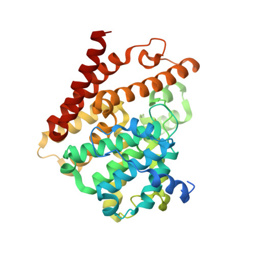 | |
UniProt | |||||
Find proteins for Q8WQX9 (Trypanosoma brucei) Explore Q8WQX9 Go to UniProtKB: Q8WQX9 | |||||
Entity Groups | |||||
| Sequence Clusters | 30% Identity50% Identity70% Identity90% Identity95% Identity100% Identity | ||||
| UniProt Group | Q8WQX9 | ||||
Sequence AnnotationsExpand | |||||
| |||||
| Ligands 5 Unique | |||||
|---|---|---|---|---|---|
| ID | Chains | Name / Formula / InChI Key | 2D Diagram | 3D Interactions | |
| D62 Query on D62 | C [auth A], H [auth B] | (4aS,8aR)-2-(1-{2-aminothieno[2,3-d]pyrimidin-4-yl}piperidin-4-yl)-4-(3,4- dimethoxyphenyl)-1,2,4a,5,8,8a-hexahydrophthalazin-1-one C27 H30 N6 O3 S VRSCGUCAJHMOSB-RBUKOAKNSA-N |  | ||
| GOL Query on GOL | G [auth A] | GLYCEROL C3 H8 O3 PEDCQBHIVMGVHV-UHFFFAOYSA-N |  | ||
| ZN Query on ZN | D [auth A], I [auth B] | ZINC ION Zn PTFCDOFLOPIGGS-UHFFFAOYSA-N |  | ||
| GAI Query on GAI | F [auth A] | GUANIDINE C H5 N3 ZRALSGWEFCBTJO-UHFFFAOYSA-N |  | ||
| MG Query on MG | E [auth A], J [auth B] | MAGNESIUM ION Mg JLVVSXFLKOJNIY-UHFFFAOYSA-N |  | ||
| Length ( Å ) | Angle ( ˚ ) |
|---|---|
| a = 111.69 | α = 90 |
| b = 119.26 | β = 108.38 |
| c = 67.97 | γ = 90 |
| Software Name | Purpose |
|---|---|
| REFMAC | refinement |
| XDS | data reduction |
| Aimless | data scaling |
| PHASER | phasing |
RCSB PDB (citation) is hosted by
RCSB PDB is a member of the

