6F6E

R2-like ligand-binding oxidase V72A mutant with anaerobically reconstituted Mn/Fe cofactor


Experimental Data Snapshot

  • Method: X-RAY DIFFRACTION
  • Resolution: 1.63 Å
  • R-Value Free: 0.201 
  • R-Value Work: 0.175 
  • R-Value Observed: 0.176 

wwPDB Validation   3D Report Full Report


Ligand Structure Quality Assessment 


This is version 1.3 of the entry. See complete history


Literature

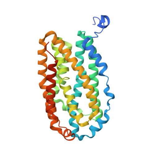
Ether cross-link formation in the R2-like ligand-binding oxidase.

Griese, J.J.Branca, R.M.M.Srinivas, V.Hogbom, M.

(2018) J Biol Inorg Chem 23: 879-886

  • DOI: https://doi.org/10.1007/s00775-018-1583-3
  • Primary Citation of Related Structures:  
    6F6C, 6F6E, 6F6F, 6F6G, 6F6H, 6F6K

  • PubMed Abstract: 

    R2-like ligand-binding oxidases contain a dinuclear metal cofactor which can consist either of two iron ions or one manganese and one iron ion, but the heterodinuclear Mn/Fe cofactor is the preferred assembly in the presence of Mn II and Fe II in vitro. We have previously shown that both types of cofactor are capable of catalyzing formation of a tyrosine-valine ether cross-link in the protein scaffold. Here we demonstrate that Mn/Fe centers catalyze cross-link formation more efficiently than Fe/Fe centers, indicating that the heterodinuclear cofactor is the biologically relevant one. We further explore the chemical potential of the Mn/Fe cofactor by introducing mutations at the cross-linking valine residue. We find that cross-link formation is possible also to the tertiary beta-carbon in an isoleucine, but not to the secondary beta-carbon or tertiary gamma-carbon in a leucine, nor to the primary beta-carbon of an alanine. These results illustrate that the reactivity of the cofactor is highly specific and directed.


  • Organizational Affiliation

    Department of Biochemistry and Biophysics, Stockholm University, 106 91, Stockholm, Sweden. julia.griese@icm.uu.se.


Macromolecules
Find similar proteins by:  (by identity cutoff)  |  3D Structure
Entity ID: 1
MoleculeChains Sequence LengthOrganismDetailsImage
Ribonucleotide reductase small subunit
A, B
316Geobacillus kaustophilus HTA426Mutation(s): 1 
Gene Names: GK2771
EC: 1.17.4.1
UniProt
Find proteins for Q5KW80 (Geobacillus kaustophilus (strain HTA426))
Explore Q5KW80 
Go to UniProtKB:  Q5KW80
Entity Groups  
Sequence Clusters30% Identity50% Identity70% Identity90% Identity95% Identity100% Identity
UniProt GroupQ5KW80
Sequence Annotations
Expand
  • Reference Sequence
Small Molecules
Experimental Data & Validation

Experimental Data

  • Method: X-RAY DIFFRACTION
  • Resolution: 1.63 Å
  • R-Value Free: 0.201 
  • R-Value Work: 0.175 
  • R-Value Observed: 0.176 
  • Space Group: C 1 2 1
Unit Cell:
Length ( Å )Angle ( ˚ )
a = 161.516α = 90
b = 55.654β = 114.11
c = 70.054γ = 90
Software Package:
Software NamePurpose
PHENIXrefinement
XDSdata reduction
XDSdata scaling
PHASERphasing

Structure Validation

View Full Validation Report


Ligand Structure Quality Assessment 


Entry History 

Deposition Data

Revision History  (Full details and data files)

  • Version 1.0: 2018-07-04
    Type: Initial release
  • Version 1.1: 2018-07-11
    Changes: Data collection, Database references
  • Version 1.2: 2018-08-01
    Changes: Data collection, Database references
  • Version 1.3: 2024-01-17
    Changes: Data collection, Database references, Derived calculations, Refinement description