6F5B

Structure of ARTD2/PARP2 WGR domain bound to double stranded DNA with 5'phosphate


Experimental Data Snapshot

  • Method: X-RAY DIFFRACTION
  • Resolution: 2.80 Å
  • R-Value Free: 0.274 
  • R-Value Work: 0.208 
  • R-Value Observed: 0.211 

wwPDB Validation   3D Report Full Report


This is version 1.4 of the entry. See complete history


Literature

Structural basis for DNA break recognition by ARTD2/PARP2.

Obaji, E.Haikarainen, T.Lehtio, L.

(2018) Nucleic Acids Res 46: 12154-12165

  • DOI: https://doi.org/10.1093/nar/gky927
  • Primary Citation of Related Structures:  
    6F1K, 6F5B, 6F5F

  • PubMed Abstract: 

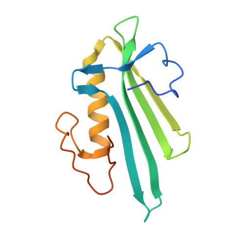
    Human ARTD2 (or PARP2) is an ADP-ribosyltransferase, which is catalytically activated by binding to damaged DNA. ARTD2 subsequently ADP-ribosylates itself and other proteins, initiating a cascade of events leading to DNA repair. In contrast to ARTD1, the founding member of the enzyme family, ARTD2 does not have specialized zinc-fingers for detecting DNA damage. The domain organization of ARTD2 includes disordered N-terminus, WGR and catalytic domains. However, the N-terminus of ARTD2 is not strictly required for the DNA dependent activity. While it is known that ARTD2 requires the WGR domain for efficient DNA binding and subsequent catalytic activation, the mechanism of DNA damage detection and subsequent catalytic activation are not completely understood. Here, we report crystal structures of ARTD2 WGR domain bound to double-strand break mimicking DNA oligonucleotides. Notably, the crystal structures revealed DNA binding mode of ARTD2 involving DNA end to end interaction. Structures demonstrate how ARTD2 recognizes nicked DNA, how it interacts with the 5'-phosphate group, and how it can mediate joining of DNA ends in vitro. Extensive mutagenesis of the ARTD2-DNA interface combined with activity, binding, and stoichiometry measurements demonstrate that the WGR domain is the key for DNA break detection.


  • Organizational Affiliation

    Faculty of Biochemistry and Molecular Medicine and Biocenter Oulu, University of Oulu, Oulu, Finland.


Macromolecules

Find similar proteins by:  (by identity cutoff)  |  3D Structure
Entity ID: 1
MoleculeChains Sequence LengthOrganismDetailsImage
Poly [ADP-ribose] polymerase 2
A, B
131Homo sapiensMutation(s): 0 
Gene Names: PARP2ADPRT2ADPRTL2
EC: 2.4.2.30
UniProt & NIH Common Fund Data Resources
Find proteins for Q9UGN5 (Homo sapiens)
Explore Q9UGN5 
Go to UniProtKB:  Q9UGN5
PHAROS:  Q9UGN5
GTEx:  ENSG00000129484 
Entity Groups  
Sequence Clusters30% Identity50% Identity70% Identity90% Identity95% Identity100% Identity
UniProt GroupQ9UGN5
Sequence Annotations
Expand
  • Reference Sequence

Find similar nucleic acids by:  Sequence   |   3D Structure  

Entity ID: 2
MoleculeChains LengthOrganismImage
DNA (5'-D(P*GP*CP*CP*TP*AP*TP*AP*GP*GP*C)-3')
C, D, E, F
10Homo sapiens
Sequence Annotations
Expand
  • Reference Sequence
Experimental Data & Validation

Experimental Data

  • Method: X-RAY DIFFRACTION
  • Resolution: 2.80 Å
  • R-Value Free: 0.274 
  • R-Value Work: 0.208 
  • R-Value Observed: 0.211 
  • Space Group: P 21 21 21
Unit Cell:
Length ( Å )Angle ( ˚ )
a = 63.16α = 90
b = 84.36β = 90
c = 95.96γ = 90
Software Package:
Software NamePurpose
REFMACrefinement
XDSdata reduction
XSCALEdata scaling
PHASERphasing

Structure Validation

View Full Validation Report


Entry History & Funding Information

Deposition Data


Funding OrganizationLocationGrant Number
Academy of FinlandFinland--
Jane and Aatos Erkko FoundationFinland--
Biocenter Oulu, University of OuluFinland--

Revision History  (Full details and data files)

  • Version 1.0: 2018-10-10
    Type: Initial release
  • Version 1.1: 2018-10-24
    Changes: Data collection, Database references
  • Version 1.2: 2018-12-26
    Changes: Data collection, Database references
  • Version 1.3: 2019-10-16
    Changes: Author supporting evidence, Data collection
  • Version 1.4: 2024-05-01
    Changes: Data collection, Database references, Refinement description