6ET6

Crystal structure of muramidase from Acinetobacter baumannii AB 5075UW prophage


Experimental Data Snapshot

  • Method: X-RAY DIFFRACTION
  • Resolution: 1.20 Å
  • R-Value Free: 0.169 
  • R-Value Work: 0.147 
  • R-Value Observed: 0.148 

wwPDB Validation   3D Report Full Report


This is version 1.1 of the entry. See complete history


Literature

Structure of anAcinetobacterBroad-Range Prophage Endolysin Reveals a C-Terminal alpha-Helix with the Proposed Role in Activity against Live Bacterial Cells.

Sykilinda, N.N.Nikolaeva, A.Y.Shneider, M.M.Mishkin, D.V.Patutin, A.A.Popov, V.O.Boyko, K.M.Klyachko, N.L.Miroshnikov, K.A.

(2018) Viruses 10

  • DOI: https://doi.org/10.3390/v10060309
  • Primary Citation of Related Structures:  
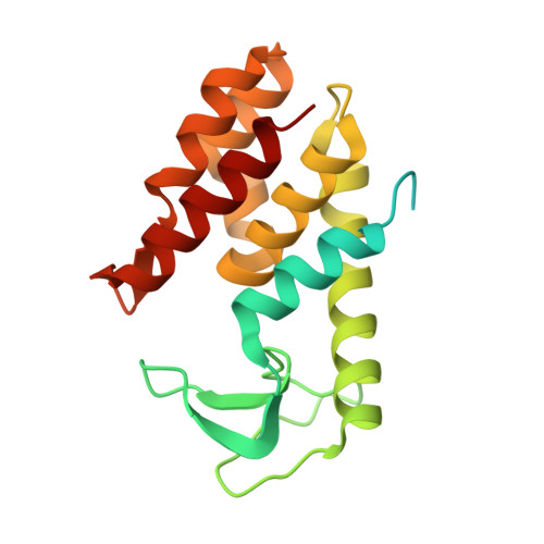
    6ET6

  • PubMed Abstract: 

    Proteins that include enzymatic domain degrading the bacterial cell wall and a domain providing transport through the bacterial outer membrane are considered as prospective compounds to combat pathogenic Gram-negative bacteria. This paper presents an isolation and study of an enzyme of this class naturally encoded in the prophage region of Acinetobacter baumannii AB 5075 genome. Recombinant protein expressed in E. coli exhibits an antimicrobial activity with respect to live cultures of Gram-negative bacteria reducing the population of viable bacteria by 1.5⁻2 log colony forming units (CFU)/mL. However the protein becomes rapidly inactivated and enables the bacteria to restore the population. AcLys structure determined by X-ray crystallography reveals a predominantly α—helical fold similar to bacteriophage P22 lysozyme. The С-terminal part of AcLys polypeptide chains forms an α—helix enriched by Lys and Arg residues exposed outside of the protein globule. Presumably this type of structure of the C-terminal α—helix has evolved evolutionally enabling the endolysin to pass the inner membrane during the host lysis or, potentially, to penetrate the outer membrane of the Gram-negative bacteria.


  • Organizational Affiliation

    Shemyakin-Ovchinnikov Institute of Bioorganic Chemistry, Russian Academy of Sciences, Moscow 117997, Russia. sykilinda@mail.ru.


Macromolecules
Find similar proteins by:  (by identity cutoff)  |  3D Structure
Entity ID: 1
MoleculeChains Sequence LengthOrganismDetailsImage
Lysozyme196Acinetobacter baumanniiMutation(s): 0 
Gene Names: rrrDABUW_1326CAS84_17450CBI29_02574
EC: 3.2.1.17
UniProt
Find proteins for A0A0D5YGK2 (Acinetobacter baumannii)
Explore A0A0D5YGK2 
Go to UniProtKB:  A0A0D5YGK2
Entity Groups  
Sequence Clusters30% Identity50% Identity70% Identity90% Identity95% Identity100% Identity
UniProt GroupA0A0D5YGK2
Sequence Annotations
Expand
  • Reference Sequence
Experimental Data & Validation

Experimental Data

  • Method: X-RAY DIFFRACTION
  • Resolution: 1.20 Å
  • R-Value Free: 0.169 
  • R-Value Work: 0.147 
  • R-Value Observed: 0.148 
  • Space Group: P 2 21 21
Unit Cell:
Length ( Å )Angle ( ˚ )
a = 30.98α = 90
b = 67β = 90
c = 75.76γ = 90
Software Package:
Software NamePurpose
HKL-2000data collection
Aimlessdata scaling
MoRDamodel building
REFMACrefinement
PDB_EXTRACTdata extraction
MOSFLMdata reduction
MOLREPphasing

Structure Validation

View Full Validation Report


Entry History & Funding Information

Deposition Data


Funding OrganizationLocationGrant Number
Russian Science Foundation GrantRussian Federation14-24-00172
Russian Federal Space Agency (Roscosmos)Russian Federation--

Revision History  (Full details and data files)

  • Version 1.0: 2018-09-05
    Type: Initial release
  • Version 1.1: 2024-01-17
    Changes: Data collection, Database references, Refinement description