


Crystal structure of KDM5B in complex with KDOPZ48a.
srikannathasan, v.To be published.
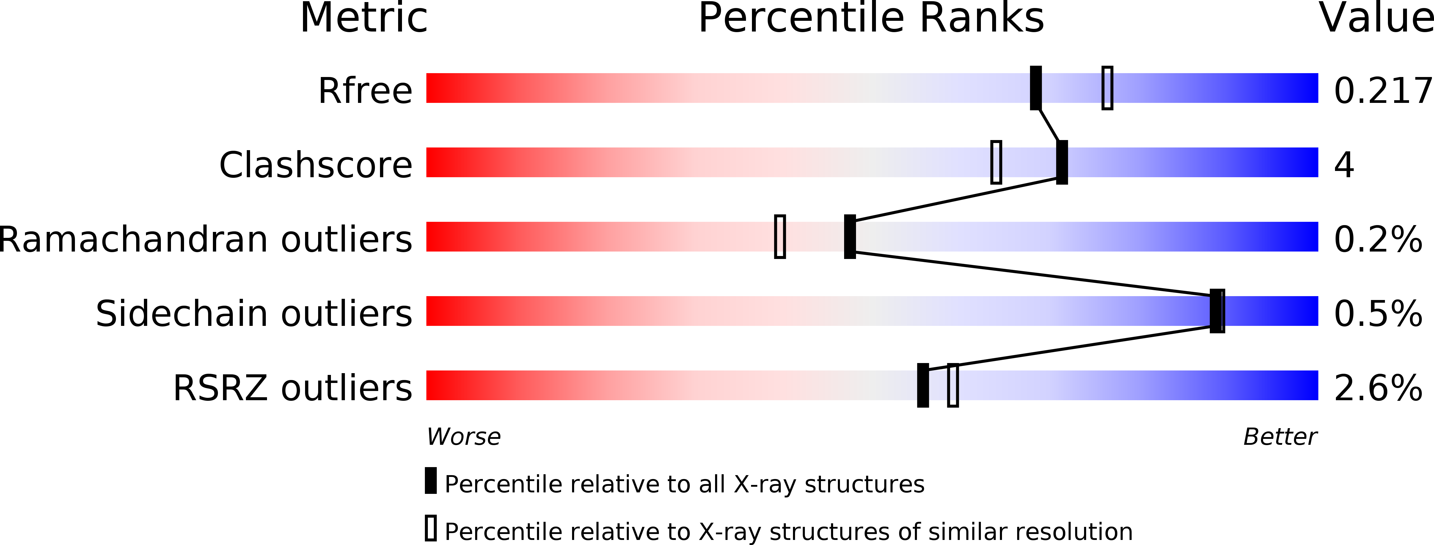
Experimental Data Snapshot
Entity ID: 1 | |||||
|---|---|---|---|---|---|
| Molecule | Chains | Sequence Length | Organism | Details | Image |
| Lysine-specific demethylase 5B,Lysine-specific demethylase 5B | 459 | Homo sapiens | Mutation(s): 0 Gene Names: KDM5B, JARID1B, PLU1, RBBP2H1 EC: 1.14.11 |  | |
UniProt & NIH Common Fund Data Resources | |||||
Find proteins for Q9UGL1 (Homo sapiens) Explore Q9UGL1 Go to UniProtKB: Q9UGL1 | |||||
PHAROS: Q9UGL1 GTEx: ENSG00000117139 | |||||
Entity Groups | |||||
| Sequence Clusters | 30% Identity50% Identity70% Identity90% Identity95% Identity100% Identity | ||||
| UniProt Group | Q9UGL1 | ||||
Sequence AnnotationsExpand | |||||
| |||||
| Ligands 5 Unique | |||||
|---|---|---|---|---|---|
| ID | Chains | Name / Formula / InChI Key | 2D Diagram | 3D Interactions | |
| B7Q (Subject of Investigation/LOI) Query on B7Q | H [auth A] | 5-[1-[1-(2-chloranylethanoyl)piperidin-4-yl]pyrazol-4-yl]-7-oxidanylidene-6-propan-2-yl-4~{H}-pyrazolo[1,5-a]pyrimidine-3-carbonitrile C20 H22 Cl N7 O2 UFTLGLOWZBFFND-UHFFFAOYSA-N |  | ||
| DMS Query on DMS | E [auth A], F [auth A], G [auth A] | DIMETHYL SULFOXIDE C2 H6 O S IAZDPXIOMUYVGZ-UHFFFAOYSA-N |  | ||
| ZN Query on ZN | B [auth A] | ZINC ION Zn PTFCDOFLOPIGGS-UHFFFAOYSA-N |  | ||
| EDO Query on EDO | I [auth A], J [auth A] | 1,2-ETHANEDIOL C2 H6 O2 LYCAIKOWRPUZTN-UHFFFAOYSA-N |  | ||
| MN Query on MN | C [auth A], D [auth A] | MANGANESE (II) ION Mn WAEMQWOKJMHJLA-UHFFFAOYSA-N |  | ||
| Length ( Å ) | Angle ( ˚ ) |
|---|---|
| a = 143.21 | α = 90 |
| b = 143.21 | β = 90 |
| c = 153.82 | γ = 120 |
| Software Name | Purpose |
|---|---|
| PHENIX | refinement |
| PDB_EXTRACT | data extraction |
| xia2 | data reduction |
| xia2 | data scaling |
| MOLREP | phasing |
RCSB PDB (citation) is hosted by
RCSB PDB is a member of the

