6DYX

Structure of VHH R419 isolated from a pre-immune phage display library


Experimental Data Snapshot

  • Method: X-RAY DIFFRACTION
  • Resolution: 1.50 Å
  • R-Value Free: 0.190 
  • R-Value Work: 0.176 
  • R-Value Observed: 0.177 

wwPDB Validation   3D Report Full Report


This is version 1.2 of the entry. See complete history


Literature

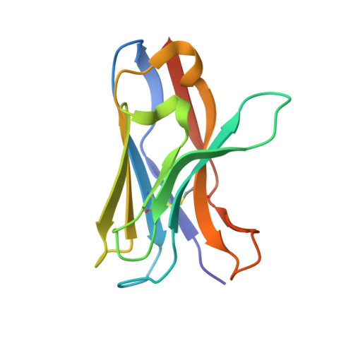
Structure of a VHH isolated from a naive phage display library.

White, B.Huh, I.Brooks, C.L.

(2019) BMC Res Notes 12: 154-154

  • DOI: https://doi.org/10.1186/s13104-019-4197-0
  • Primary Citation of Related Structures:  
    6DYX

  • PubMed Abstract: 

    To determine the X-ray structure and biophysical properties of a Camelid V H H isolated from a naïve phage display library. Single domain antibodies (V H H) derived from the unique immune system of the Camelidae family have gained traction as useful tools for biotechnology as well as a source of potentially novel therapeutics. Here we report the structure and biophysical characterization of a V H H originally isolated from a naïve camelid phage display library. V H H R419 has a melting temperate of 66 °C and was found to be a monomer in solution. The protein crystallized in space group P6 5 22 and the structure was solved by molecular replacement to a resolution of 1.5 Å. The structure revealed a flat paratope with CDR loops that could be classified into existing canonical loop structures. A combination of high expression yield, stability and rapid crystallization might make R419 into a candidate scaffold for CDR grafting and homology modeling.


  • Organizational Affiliation

    Department of Chemistry, California State University Fresno, 2555 E San Ramon Ave, Fresno, CA, 93740, USA.


Macromolecules
Find similar proteins by:  (by identity cutoff)  |  3D Structure
Entity ID: 1
MoleculeChains Sequence LengthOrganismDetailsImage
VHH R419A [auth D]124Vicugna pacosMutation(s): 0 
Entity Groups  
Sequence Clusters30% Identity50% Identity70% Identity90% Identity95% Identity100% Identity
Sequence Annotations
Expand
  • Reference Sequence
Experimental Data & Validation

Experimental Data

  • Method: X-RAY DIFFRACTION
  • Resolution: 1.50 Å
  • R-Value Free: 0.190 
  • R-Value Work: 0.176 
  • R-Value Observed: 0.177 
  • Space Group: P 65 2 2
Unit Cell:
Length ( Å )Angle ( ˚ )
a = 58.281α = 90
b = 58.281β = 90
c = 155.358γ = 120
Software Package:
Software NamePurpose
PHENIXrefinement
SCALAdata scaling
PDB_EXTRACTdata extraction
xia2data reduction
PHASERphasing

Structure Validation

View Full Validation Report


Entry History & Funding Information

Deposition Data

  • Released Date: 2019-04-03 
  • Deposition Author(s): Brooks, C.L.

Funding OrganizationLocationGrant Number
National Institutes of Health/National Institute of General Medical Sciences (NIH/NIGMS)United StatesSC3GM112532

Revision History  (Full details and data files)

  • Version 1.0: 2019-04-03
    Type: Initial release
  • Version 1.1: 2020-01-01
    Changes: Author supporting evidence
  • Version 1.2: 2023-10-11
    Changes: Data collection, Database references, Refinement description