6DUQ

Structure of a Rho-NusG KOW domain complex


Experimental Data Snapshot

  • Method: X-RAY DIFFRACTION
  • Resolution: 3.70 Å
  • R-Value Free: 0.300 
  • R-Value Work: 0.287 
  • R-Value Observed: 0.288 

wwPDB Validation   3D Report Full Report


Ligand Structure Quality Assessment 


This is version 1.5 of the entry. See complete history


Literature

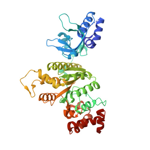
Mechanism for the Regulated Control of Bacterial Transcription Termination by a Universal Adaptor Protein.

Lawson, M.R.Ma, W.Bellecourt, M.J.Artsimovitch, I.Martin, A.Landick, R.Schulten, K.Berger, J.M.

(2018) Mol Cell 71: 911-922.e4

  • DOI: https://doi.org/10.1016/j.molcel.2018.07.014
  • Primary Citation of Related Structures:  
    6DUQ

  • PubMed Abstract: 

    NusG/Spt5 proteins are the only transcription factors utilized by all cellular organisms. In enterobacteria, NusG antagonizes the transcription termination activity of Rho, a hexameric helicase, during the synthesis of ribosomal and actively translated mRNAs. Paradoxically, NusG helps Rho act on untranslated transcripts, including non-canonical antisense RNAs and those arising from translational stress; how NusG fulfills these disparate functions is unknown. Here, we demonstrate that NusG activates Rho by assisting helicase isomerization from an open-ring, RNA-loading state to a closed-ring, catalytically active translocase. A crystal structure of closed-ring Rho in complex with NusG reveals the physical basis for this activation and further explains how Rho is excluded from translationally competent RNAs. This study demonstrates how a universally conserved transcription factor acts to modulate the activity of a ring-shaped ATPase motor and establishes how the innate sequence bias of a termination factor can be modulated to silence pervasive, aberrant transcription.


  • Organizational Affiliation

    Department of Molecular and Cell Biology, University of California at Berkeley, Berkeley, CA 94720, USA.


Macromolecules

Find similar proteins by:  (by identity cutoff)  |  3D Structure
Entity ID: 1
MoleculeChains Sequence LengthOrganismDetailsImage
Transcription termination factor Rho422Escherichia coli M718Mutation(s): 0 
Gene Names: rhoECJG_05123
EC: 3.6.4
UniProt
Find proteins for P0AG30 (Escherichia coli (strain K12))
Explore P0AG30 
Go to UniProtKB:  P0AG30
Entity Groups  
Sequence Clusters30% Identity50% Identity70% Identity90% Identity95% Identity100% Identity
UniProt GroupP0AG30
Sequence Annotations
Expand
  • Reference Sequence
Find similar proteins by:  (by identity cutoff)  |  3D Structure
Entity ID: 2
MoleculeChains Sequence LengthOrganismDetailsImage
Transcription termination/antitermination protein NusG
M, N, O, P, Q
M, N, O, P, Q, R, S, T, U, V, W, X
60Escherichia coli M605Mutation(s): 0 
Gene Names: nusGECIG_05396
UniProt
Find proteins for P0AFG0 (Escherichia coli (strain K12))
Explore P0AFG0 
Go to UniProtKB:  P0AFG0
Entity Groups  
Sequence Clusters30% Identity50% Identity70% Identity90% Identity95% Identity100% Identity
UniProt GroupP0AFG0
Sequence Annotations
Expand
  • Reference Sequence

Find similar nucleic acids by:  Sequence   |   3D Structure  

Entity ID: 3
MoleculeChains LengthOrganismImage
rU12
Y, Z
12synthetic construct
Sequence Annotations
Expand
  • Reference Sequence
Small Molecules
Ligands 3 Unique
IDChains Name / Formula / InChI Key2D Diagram3D Interactions
ADP
Query on ADP

Download Ideal Coordinates CCD File 
AA [auth L]
BB [auth F]
DA [auth A]
EB [auth J]
GA [auth G]
AA [auth L],
BB [auth F],
DA [auth A],
EB [auth J],
GA [auth G],
HB [auth E],
JA [auth B],
MA [auth H],
PA [auth C],
SA [auth I],
VA [auth D],
YA [auth K]
ADENOSINE-5'-DIPHOSPHATE
C10 H15 N5 O10 P2
XTWYTFMLZFPYCI-KQYNXXCUSA-N
BEF
Query on BEF

Download Ideal Coordinates CCD File 
AB [auth K]
CA [auth L]
DB [auth F]
FA [auth A]
GB [auth J]
AB [auth K],
CA [auth L],
DB [auth F],
FA [auth A],
GB [auth J],
IA [auth G],
JB [auth E],
LA [auth B],
OA [auth H],
RA [auth C],
UA [auth I],
XA [auth D]
BERYLLIUM TRIFLUORIDE ION
Be F3
OGIAHMCCNXDTIE-UHFFFAOYSA-K
MG
Query on MG

Download Ideal Coordinates CCD File 
BA [auth L]
CB [auth F]
EA [auth A]
FB [auth J]
HA [auth G]
BA [auth L],
CB [auth F],
EA [auth A],
FB [auth J],
HA [auth G],
IB [auth E],
KA [auth B],
NA [auth H],
QA [auth C],
TA [auth I],
WA [auth D],
ZA [auth K]
MAGNESIUM ION
Mg
JLVVSXFLKOJNIY-UHFFFAOYSA-N
Experimental Data & Validation

Experimental Data

  • Method: X-RAY DIFFRACTION
  • Resolution: 3.70 Å
  • R-Value Free: 0.300 
  • R-Value Work: 0.287 
  • R-Value Observed: 0.288 
  • Space Group: P 1
Unit Cell:
Length ( Å )Angle ( ˚ )
a = 126.35α = 89.94
b = 126.32β = 89.9
c = 166.82γ = 60.01
Software Package:
Software NamePurpose
PHENIXrefinement
XDSdata reduction
XSCALEdata scaling
PHASERphasing

Structure Validation

View Full Validation Report


Ligand Structure Quality Assessment 


Entry History & Funding Information

Deposition Data


Funding OrganizationLocationGrant Number
National Institutes of Health/National Institute of General Medical Sciences (NIH/NIGMS)United StatesGM071747
Other privateUnited StatesG. Harold and Leila Y. Mathers Foundation

Revision History  (Full details and data files)

  • Version 1.0: 2018-09-05
    Type: Initial release
  • Version 1.1: 2018-10-03
    Changes: Data collection, Database references
  • Version 1.2: 2019-04-17
    Changes: Author supporting evidence, Data collection
  • Version 1.3: 2020-01-01
    Changes: Author supporting evidence
  • Version 1.4: 2020-01-29
    Changes: Derived calculations
  • Version 1.5: 2024-03-13
    Changes: Data collection, Database references, Derived calculations