Contribution of an unusual CDR2 element of a single domain antibody in ricin toxin binding affinity and neutralizing activity.
Rudolph, M.J., Vance, D.J., Kelow, S., Angalakurthi, S.K., Nguyen, S., Davis, S.A., Rong, Y., Middaugh, C.R., Weis, D.D., Dunbrack Jr., R., Karanicolas, J., Mantis, N.J.(2018) Protein Eng Des Sel 31: 277-287
- PubMed: 30265352
- DOI: https://doi.org/10.1093/protein/gzy022
- Primary Citation of Related Structures:
6CWG, 6CWK - PubMed Abstract:
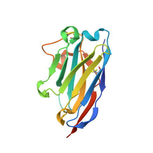
Ricin toxin's enzymatic subunit (RTA) has been subjected to intensive B cell epitope mapping studies using a combination of competition ELISAs, hydrogen exchange-mass spectrometry and X-ray crystallography. Those studies identified four spatially distinct clusters (I-IV) of toxin-neutralizing epitopes on the surface of RTA. Here we describe A9, a new single domain camelid antibody (VHH) that was proposed to recognize a novel epitope on RTA that straddles clusters I and III. The X-ray crystal structure of A9 bound to RTA (2.6 Å resolution) revealed extensive antibody contact with RTA's β-strand h (732 Å2 buried surface area; BSA), along with limited engagement with α-helix D (90 Å2) and α-helix C (138 Å2). Collectively, these contacts explain the overlap between epitope clusters I and III, as identified by competition ELISA. However, considerable binding affinity, and, consequently, toxin-neutralizing activity of A9 is mediated by an unusual CDR2 containing five consecutive Gly residues that interact with α-helix B (82 Å2), a known neutralizing hotspot on RTA. Removal of a single Gly residue from the penta-glycine stretch in CDR2 reduced A9's binding affinity by 10-fold and eliminated toxin-neutralizing activity. Computational modeling indicates that removal of a Gly from CDR2 does not perturb contact with RTA per se, but results in the loss of an intramolecular hydrogen bond network involved in stabilizing CDR2 in the unbound state. These results reveal a novel configuration of a CDR2 element involved in neutralizing ricin toxin.
Organizational Affiliation:
New York Structural Biology Center, New York, NY, USA.