


Characteristic conformational changes on the distal and proximal surfaces of cytochrome P450 2D6 in response to substrate binding
Yang, Y.T., Fujita, F., Wang, P.F., Im, S.C., Pearl, N.M., Meagher, J., Stuckey, J., Waskell, L.To be published.
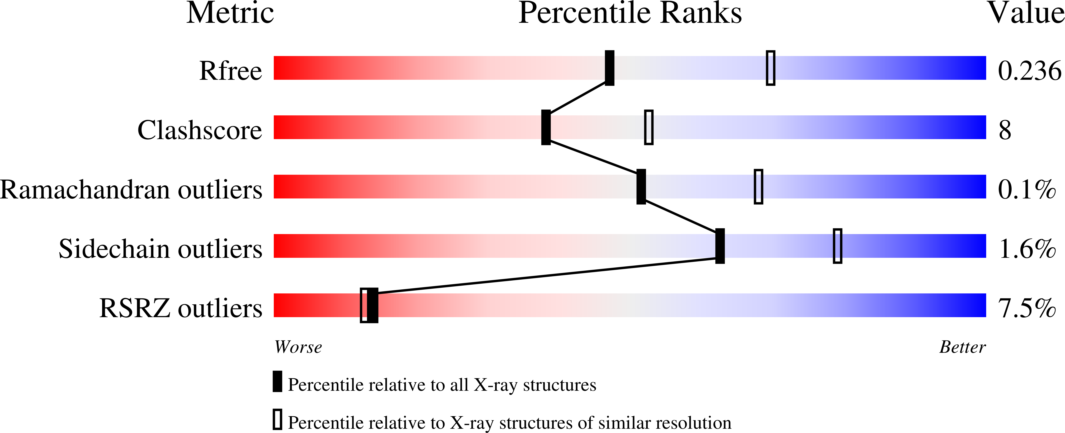
Experimental Data Snapshot
Entity ID: 1 | |||||
|---|---|---|---|---|---|
| Molecule | Chains | Sequence Length | Organism | Details | Image |
| Cytochrome P450 2D6 | 479 | Homo sapiens | Mutation(s): 1 Gene Names: CYP2D6, CYP2DL1 EC: 1.14.14.1 |  | |
UniProt & NIH Common Fund Data Resources | |||||
Find proteins for P10635 (Homo sapiens) Explore P10635 Go to UniProtKB: P10635 | |||||
PHAROS: P10635 GTEx: ENSG00000100197 | |||||
Entity Groups | |||||
| Sequence Clusters | 30% Identity50% Identity70% Identity90% Identity95% Identity100% Identity | ||||
| UniProt Group | P10635 | ||||
Sequence AnnotationsExpand | |||||
| |||||
| Ligands 4 Unique | |||||
|---|---|---|---|---|---|
| ID | Chains | Name / Formula / InChI Key | 2D Diagram | 3D Interactions | |
| HEM Query on HEM | C [auth A], I [auth B] | PROTOPORPHYRIN IX CONTAINING FE C34 H32 Fe N4 O4 KABFMIBPWCXCRK-RGGAHWMASA-L |  | ||
| CPS Query on CPS | E [auth A], K [auth B] | 3-[(3-CHOLAMIDOPROPYL)DIMETHYLAMMONIO]-1-PROPANESULFONATE C32 H58 N2 O7 S UMCMPZBLKLEWAF-BCTGSCMUSA-N |  | ||
| PN0 Query on PN0 | D [auth A], J [auth B] | Prinomastat C18 H21 N3 O5 S2 YKPYIPVDTNNYCN-INIZCTEOSA-N |  | ||
| ZN Query on ZN | F [auth A] G [auth A] H [auth A] L [auth B] M [auth B] | ZINC ION Zn PTFCDOFLOPIGGS-UHFFFAOYSA-N |  | ||
| Length ( Å ) | Angle ( ˚ ) |
|---|---|
| a = 57.472 | α = 90 |
| b = 126.527 | β = 90 |
| c = 192.258 | γ = 90 |
| Software Name | Purpose |
|---|---|
| PHENIX | refinement |
| HKL-2000 | data reduction |
| HKL-2000 | data scaling |
| PHASER | phasing |
RCSB PDB (citation) is hosted by
RCSB PDB is a member of the

