6C1R

Crystal structure of human C5a receptor in complex with an orthosteric antagonist PMX53 and an allosteric antagonist avacopan


Experimental Data Snapshot

  • Method: X-RAY DIFFRACTION
  • Resolution: 2.20 Å
  • R-Value Free: 0.224 
  • R-Value Work: 0.192 
  • R-Value Observed: 0.194 

wwPDB Validation   3D Report Full Report


Ligand Structure Quality Assessment 


This is version 1.5 of the entry. See complete history


Literature

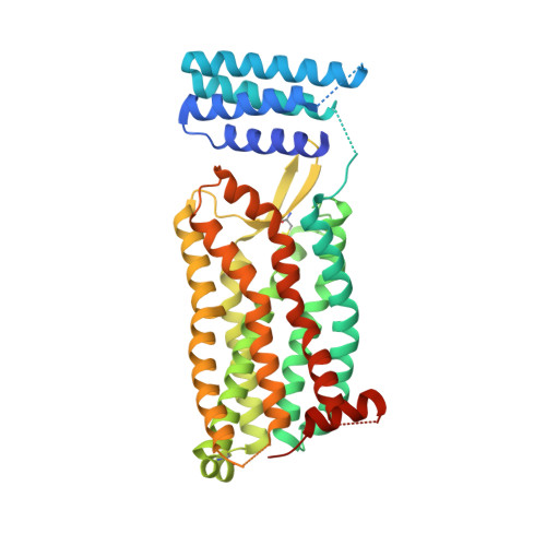
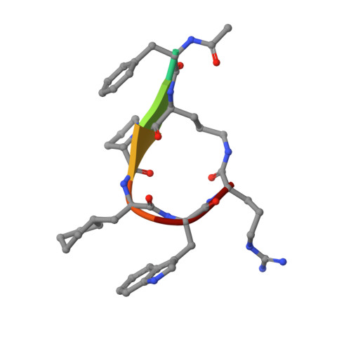
Orthosteric and allosteric action of the C5a receptor antagonists.

Liu, H.Kim, H.R.Deepak, R.N.V.K.Wang, L.Chung, K.Y.Fan, H.Wei, Z.Zhang, C.

(2018) Nat Struct Mol Biol 25: 472-481

  • DOI: https://doi.org/10.1038/s41594-018-0067-z
  • Primary Citation of Related Structures:  
    6C1Q, 6C1R

  • PubMed Abstract: 

    The C5a receptor (C5aR) is a G-protein-coupled receptor (GPCR) that can induce strong inflammatory response to the anaphylatoxin C5a. Targeting C5aR has emerged as a novel anti-inflammatory therapeutic method. However, developing potent C5aR antagonists as drugs has proven difficult. Here, we report two crystal structures of human C5aR in ternary complexes with the peptide antagonist PMX53 and a non-peptide antagonist, either avacopan or NDT9513727. The structures, together with other biophysical, computational docking and cell-based signaling data, reveal the orthosteric action of PMX53 and its effect of stabilizing the C5aR structure, as well as the allosteric action of chemically diverse non-peptide C5aR antagonists with different binding poses. Structural comparison analysis suggests the presence of similar allosteric sites in other GPCRs. We also discuss critical structural features of C5aR in activation, including a novel conformation of helix 8. On the basis of our results, we suggest novel strategies for developing C5aR-targeting drugs.


  • Organizational Affiliation

    Department of Pharmacology and Chemical Biology, School of Medicine, University of Pittsburgh, Pittsburgh, PA, USA.


Macromolecules
Find similar proteins by:  (by identity cutoff)  |  3D Structure
Entity ID: 1
MoleculeChains Sequence LengthOrganismDetailsImage
Soluble cytochrome b562, C5a anaphylatoxin chemotactic receptor 1 chimeraA [auth B]422Escherichia coliHomo sapiens
This entity is chimeric
Mutation(s): 3 
Gene Names: cybCC5AR1C5ARC5R1
Membrane Entity: Yes 
UniProt & NIH Common Fund Data Resources
Find proteins for P0ABE7 (Escherichia coli)
Explore P0ABE7 
Go to UniProtKB:  P0ABE7
Find proteins for P21730 (Homo sapiens)
Explore P21730 
Go to UniProtKB:  P21730
PHAROS:  P21730
GTEx:  ENSG00000197405 
Entity Groups  
Sequence Clusters30% Identity50% Identity70% Identity90% Identity95% Identity100% Identity
UniProt GroupsP0ABE7P21730
Sequence Annotations
Expand
  • Reference Sequence

Find similar proteins by:  Sequence   |   3D Structure  

Entity ID: 2
MoleculeChains Sequence LengthOrganismDetailsImage
PMX53B [auth L]7synthetic constructMutation(s): 0 
Sequence Annotations
Expand
  • Reference Sequence
Small Molecules
Ligands 6 Unique
IDChains Name / Formula / InChI Key2D Diagram3D Interactions
EFD
Query on EFD

Download Ideal Coordinates CCD File 
C [auth B]avacopan
C33 H35 F4 N3 O2
PUKBOVABABRILL-YZNIXAGQSA-N
OLC
Query on OLC

Download Ideal Coordinates CCD File 
H [auth B],
I [auth B]
(2R)-2,3-dihydroxypropyl (9Z)-octadec-9-enoate
C21 H40 O4
RZRNAYUHWVFMIP-GDCKJWNLSA-N
OLA
Query on OLA

Download Ideal Coordinates CCD File 
J [auth B],
K [auth B],
L [auth B],
M [auth B]
OLEIC ACID
C18 H34 O2
ZQPPMHVWECSIRJ-KTKRTIGZSA-N
PGE
Query on PGE

Download Ideal Coordinates CCD File 
G [auth B]TRIETHYLENE GLYCOL
C6 H14 O4
ZIBGPFATKBEMQZ-UHFFFAOYSA-N
MLI
Query on MLI

Download Ideal Coordinates CCD File 
D [auth B],
E [auth B]
MALONATE ION
C3 H2 O4
OFOBLEOULBTSOW-UHFFFAOYSA-L
NA
Query on NA

Download Ideal Coordinates CCD File 
F [auth B]SODIUM ION
Na
FKNQFGJONOIPTF-UHFFFAOYSA-N
Modified Residues  2 Unique
IDChains TypeFormula2D DiagramParent
YCM
Query on YCM
A [auth B]L-PEPTIDE LINKINGC5 H10 N2 O3 SCYS
ORN
Query on ORN
B [auth L]L-PEPTIDE LINKINGC5 H12 N2 O2ALA
Biologically Interesting Molecules (External Reference) 1 Unique
Experimental Data & Validation

Experimental Data

  • Method: X-RAY DIFFRACTION
  • Resolution: 2.20 Å
  • R-Value Free: 0.224 
  • R-Value Work: 0.192 
  • R-Value Observed: 0.194 
  • Space Group: P 1 21 1
Unit Cell:
Length ( Å )Angle ( ˚ )
a = 58.411α = 90
b = 52.608β = 106.08
c = 83.865γ = 90
Software Package:
Software NamePurpose
HKL-2000data reduction
HKL-2000data scaling
PHASERphasing
PHENIXrefinement
PDB_EXTRACTdata extraction

Structure Validation

View Full Validation Report


Ligand Structure Quality Assessment 


Entry History & Funding Information

Deposition Data


Funding OrganizationLocationGrant Number
National Natural Science Foundation of China (NSFC)China--

Revision History  (Full details and data files)

  • Version 1.0: 2018-05-30
    Type: Initial release
  • Version 1.1: 2018-06-20
    Changes: Data collection, Database references
  • Version 1.2: 2018-09-05
    Changes: Data collection, Structure summary
  • Version 1.3: 2020-01-08
    Changes: Author supporting evidence
  • Version 1.4: 2023-10-04
    Changes: Data collection, Database references, Derived calculations, Refinement description
  • Version 1.5: 2023-11-15
    Changes: Data collection