


Structure of 2-dehydro-3-deoxyphosphooctonate aldolase from Acinetobacter baumannii
Abendroth, J., Delker, S.L., Lorimer, D.D., Edwards, T.E.To be published.
Experimental Data Snapshot
wwPDB Validation 3D Report Full Report

Entity ID: 1 | |||||
|---|---|---|---|---|---|
| Molecule | Chains | Sequence Length | Organism | Details | Image |
| 2-dehydro-3-deoxyphosphooctonate aldolase | 289 | Acinetobacter baumannii AB307-0294 | Mutation(s): 0 Gene Names: kdsA, ABBFA_001556 EC: 2.5.1.55 | 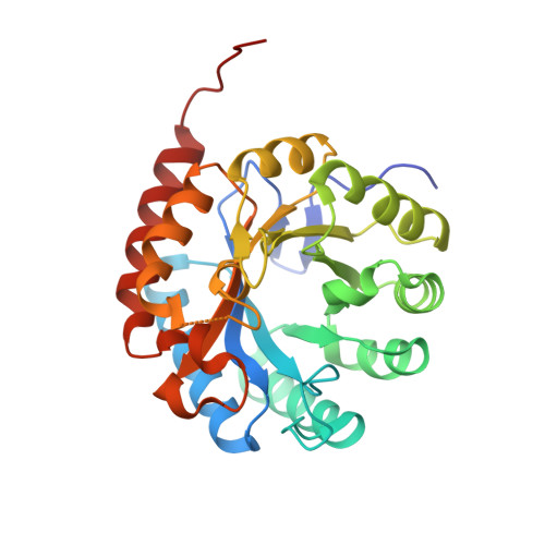 | |
UniProt | |||||
Find proteins for B7H226 (Acinetobacter baumannii (strain AB307-0294)) Explore B7H226 Go to UniProtKB: B7H226 | |||||
Entity Groups | |||||
| Sequence Clusters | 30% Identity50% Identity70% Identity90% Identity95% Identity100% Identity | ||||
| UniProt Group | B7H226 | ||||
Sequence AnnotationsExpand | |||||
| |||||
| Ligands 1 Unique | |||||
|---|---|---|---|---|---|
| ID | Chains | Name / Formula / InChI Key | 2D Diagram | 3D Interactions | |
| SO4 Query on SO4 | C [auth A], D [auth A], E [auth B], F [auth B] | SULFATE ION O4 S QAOWNCQODCNURD-UHFFFAOYSA-L |  | ||
| Length ( Å ) | Angle ( ˚ ) |
|---|---|
| a = 77.69 | α = 90 |
| b = 83.93 | β = 90 |
| c = 87.7 | γ = 90 |
| Software Name | Purpose |
|---|---|
| XSCALE | data scaling |
| PHENIX | refinement |
| PDB_EXTRACT | data extraction |
| XDS | data reduction |
| MoRDa | phasing |
| Coot | model building |
RCSB PDB (citation) is hosted by
RCSB PDB is a member of the

