6B9O

Structure of GH 38 Jack Bean alpha-mannosidase


Experimental Data Snapshot

  • Method: X-RAY DIFFRACTION
  • Resolution: 1.84 Å
  • R-Value Free: 0.216 
  • R-Value Work: 0.175 
  • R-Value Observed: 0.177 

wwPDB Validation   3D Report Full Report


This is version 2.1 of the entry. See complete history


Literature

Structural Basis of Outstanding Multivalent Effects in Jack Bean alpha-Mannosidase Inhibition.

Howard, E.Cousido-Siah, A.Lepage, M.L.Schneider, J.P.Bodlenner, A.Mitschler, A.Meli, A.Izzo, I.Alvarez, H.A.Podjarny, A.Compain, P.

(2018) Angew Chem Int Ed Engl 57: 8002-8006

  • DOI: https://doi.org/10.1002/anie.201801202
  • Primary Citation of Related Structures:  
    6B9O, 6B9P

  • PubMed Abstract: 

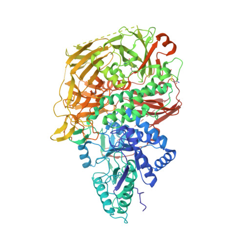
    Multivalent design of glycosidase inhibitors is a promising strategy for the treatment of diseases involving enzymatic hydrolysis of glycosidic bonds in carbohydrates. An essential prerequisite for successful applications is the atomic-level understanding of how outstanding binding enhancement occurs with multivalent inhibitors. Herein we report the first high-resolution crystal structures of the Jack bean α-mannosidase (JBα-man) in apo and inhibited states. The three-dimensional structure of JBα-man in complex with the multimeric cyclopeptoid-based inhibitor displaying the largest binding enhancements reported so far provides decisive insight into the molecular mechanisms underlying multivalent effects in glycosidase inhibition.


  • Organizational Affiliation

    Department of Integrative Biology, Institut de Génétique et de Biologie Moléculaire et Cellulaire, CNRS, INSERM, UdS, 1 rue Laurent Fries, 67404, Illkirch CEDEX, France.


Macromolecules
Find similar proteins by:  (by identity cutoff)  |  3D Structure
Entity ID: 1
MoleculeChains Sequence LengthOrganismDetailsImage
Alpha-mannosidase from Canavalia ensiformis (jack bean)
A, B
981Canavalia ensiformisMutation(s): 0 
EC: 3.2.1.24
UniProt
Find proteins for C0HJB3 (Canavalia ensiformis)
Explore C0HJB3 
Go to UniProtKB:  C0HJB3
Entity Groups  
Sequence Clusters30% Identity50% Identity70% Identity90% Identity95% Identity100% Identity
UniProt GroupC0HJB3
Sequence Annotations
Expand
  • Reference Sequence
Oligosaccharides

Help

Entity ID: 2
MoleculeChains Length2D Diagram Glycosylation3D Interactions
alpha-D-mannopyranose-(1-3)-alpha-D-mannopyranose-(1-2)-alpha-D-mannopyranose-(1-2)-alpha-D-mannopyranose-(1-3)-[alpha-D-mannopyranose-(1-2)-alpha-D-mannopyranose-(1-3)-[alpha-D-mannopyranose-(1-2)-alpha-D-mannopyranose-(1-6)]alpha-D-mannopyranose-(1-6)]beta-D-mannopyranose-(1-4)-2-acetamido-2-deoxy-beta-D-glucopyranose-(1-4)-2-acetamido-2-deoxy-beta-D-glucopyranose
C, E
12N-Glycosylation
Glycosylation Resources
GlyTouCan:  G77969OS
GlyCosmos:  G77969OS
GlyGen:  G77969OS
Entity ID: 3
MoleculeChains Length2D Diagram Glycosylation3D Interactions
2-acetamido-2-deoxy-beta-D-glucopyranose-(1-4)-2-acetamido-2-deoxy-beta-D-glucopyranose
D, F
2N-Glycosylation
Glycosylation Resources
GlyTouCan:  G42666HT
GlyCosmos:  G42666HT
GlyGen:  G42666HT
Experimental Data & Validation

Experimental Data

  • Method: X-RAY DIFFRACTION
  • Resolution: 1.84 Å
  • R-Value Free: 0.216 
  • R-Value Work: 0.175 
  • R-Value Observed: 0.177 
  • Space Group: P 21 21 21
Unit Cell:
Length ( Å )Angle ( ˚ )
a = 67.305α = 90
b = 119.667β = 90
c = 277.689γ = 90
Software Package:
Software NamePurpose
PHENIXrefinement
XDSdata reduction
XDSdata scaling
PHASERphasing

Structure Validation

View Full Validation Report


Entry History 

Deposition Data

Revision History  (Full details and data files)

  • Version 1.0: 2018-09-26
    Type: Initial release
  • Version 2.0: 2020-07-29
    Type: Remediation
    Reason: Carbohydrate remediation
    Changes: Advisory, Atomic model, Data collection, Derived calculations, Structure summary
  • Version 2.1: 2023-10-04
    Changes: Data collection, Database references, Derived calculations, Refinement description, Structure summary